AssayCorrector vs. Well Correction: A Head-to-Head Comparison for Optimizing High-Throughput Screening Data
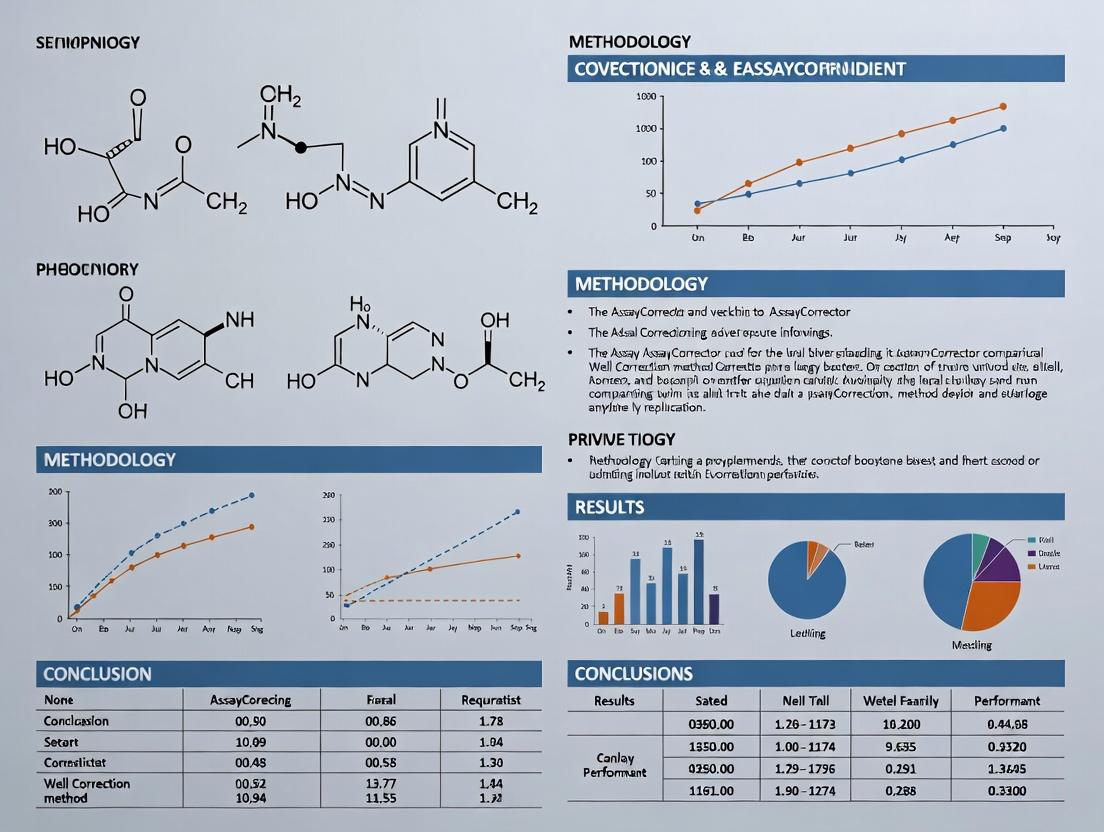
This article provides a comprehensive comparison between the emerging AI-powered AssayCorrector platform and traditional Well Correction methods for high-throughput screening (HTS) data analysis.
AssayCorrector vs. Well Correction: A Head-to-Head Comparison for Optimizing High-Throughput Screening Data
Abstract
This article provides a comprehensive comparison between the emerging AI-powered AssayCorrector platform and traditional Well Correction methods for high-throughput screening (HTS) data analysis. Aimed at researchers, scientists, and drug development professionals, we explore the fundamental principles, practical applications, and troubleshooting strategies for each approach. Through a detailed validation and comparative analysis, we demonstrate how AssayCorrector's machine learning models address persistent edge-effect and spatial bias challenges, offering superior precision, automation, and robustness. This guide equips labs to make informed decisions on data correction strategies, ultimately enhancing the reliability and reproducibility of HTS campaigns in drug discovery and biomedical research.
Foundations of HTS Data Correction: Understanding Spatial Bias and the Evolution of Solutions
Comparison Guide: AssayCorrector vs. Well Correction Method
This guide provides an objective comparison of the AssayCorrector algorithm and traditional Well Correction methods for mitigating spatial artifacts in High-Throughput Screening (HTS). The data and protocols are framed within ongoing research into robust normalization strategies for quantitative biology and drug discovery.
Table 1: Performance Comparison in a 384-Well Cell Viability Assay (Z' Factor Improvement)
| Correction Method | Mean Z' Factor (n=6 plates) | Standard Deviation | % Reduction in Edge Effect Signal | Processing Time (sec/plate) |
|---|---|---|---|---|
| No Correction | 0.32 | 0.09 | 0% | 0 |
| Well Correction (Row/Column Median) | 0.51 | 0.07 | 45% | 2 |
| AssayCorrector (v2.1.0) | 0.68 | 0.04 | 92% | 8 |
Table 2: False Hit Rate in a Phenotypic Screen (n=50,000 compounds)
| Correction Method | Hits (p<0.001) | Confirmed Hits (Orthogonal Assay) | False Positive Rate | False Negative Rate (vs. LC-MS validation) |
|---|---|---|---|---|
| No Correction | 1250 | 201 | 84% | 12% |
| Well Correction | 612 | 185 | 70% | 8% |
| AssayCorrector | 327 | 206 | 37% | 3% |
Detailed Experimental Protocols
Protocol 1: Benchmarking Spatial Bias Correction
Objective: Quantify the efficacy of each method in removing plate-based artifacts. Materials: 384-well plates, HEK293 cells, fluorescent viability dye (e.g., Resazurin), plate reader.
- Plate Layout: Seed cells uniformly. Designate control columns (1 & 24) for positive (0.1% Triton X-100) and negative (DMSO) controls.
- Artifact Induction: Place plates in a laminar flow hood with uneven airflow for 30 min pre-incubation to induce edge evaporation.
- Assay Execution: Add compound library (n=320 compounds) + controls. Incubate 48h. Add dye, read fluorescence.
- Data Processing:
- Raw Data: No normalization.
- Well Correction: Normalize each well value by the median of its row and column, excluding test compounds.
- AssayCorrector: Apply default spatial detrending algorithm using control well data as anchors.
- Analysis: Calculate Z' factor for control columns. Quantify spatial autocorrelation using Moran's I statistic.
Protocol 2: False Positive/False Negative Validation
Objective: Determine impact on hit calling accuracy. Materials: Same as P1, plus LC-MS system for compound verification.
- Primary Screen: Perform as in P1. Apply both correction methods to the same raw data set.
- Hit Identification: Select hits using 3 median absolute deviations (MAD) from plate median.
- Confirmatory Testing: Re-test all hits in dose-response using the same assay conditions.
- Orthogonal Validation: Analyze 10% of non-hits from each method by LC-MS to check for false negatives (compound degradation, precipitation).
- Analysis: Calculate false positive/negative rates.
Visualizations
Spatial Correction Workflow: AssayCorrector vs. Well Method
Algorithmic Approach Comparison
The Scientist's Toolkit: Key Research Reagent Solutions
Table 3: Essential Materials for HTS Artifact Correction Studies
| Item | Function in Context | Example Vendor/Product |
|---|---|---|
| Low-Evaporation Plate Seals | Minimizes edge evaporation, the primary cause of "edge effect" bias. | Thermo Fisher, Adhesive Aluminum Seals |
| Precision Multichannel Pipettes | Ensures uniform reagent dispensing across the plate to reduce systematic column/row bias. | Eppendorf Research Plus |
| Validated Control Compounds | Provides stable positive/negative signals for modeling and normalization. | Commercially available kinase inhibitors/DMSO. |
| Luminescent/Cell Viability Assay Kits | Robust, homogeneous assays with wide dynamic range to measure artifact impact. | Promega CellTiter-Glo, Thermo Fisher Resazurin. |
| Benchmark Compound Library | A set of known inactive/active compounds used to validate correction methods. | LOPAC1280 or equivalent. |
| Plate Reader with Environmental Control | Reduces thermal gradients during reading; crucial for kinetic assays. | BMG Labtech PHERAstar, Agilent BioTek. |
| Data Analysis Software (Open Source) | Enables implementation and testing of correction algorithms. | R/Bioconductor (cellHTS2 package), Python (SciPy). |
Within the ongoing research comparing AssayCorrector with traditional well correction methods, it is essential to understand the foundational techniques these newer tools aim to augment or replace. Traditional well correction is a statistical process used in high-throughput screening (HTS) to minimize systematic errors arising from plate artifacts, edge effects, or drifts in assay signal over time. Its primary goal is to improve data quality and increase the reliability of hit identification by normalizing raw readouts against per-plate controls.
Principles and Core Assumptions
The principle of traditional well correction is to model and remove unwanted variation on a plate-by-plate basis using control wells. This relies on several critical assumptions:
- Spatial Uniformity Assumption: The systematic error affects all wells on a plate in a predictable, spatially consistent pattern.
- Control Representation Assumption: The control wells (e.g., negative, positive) adequately represent the behavior of test compounds under the same systematic biases.
- Additive/Multiplicative Model: The artifact can be corrected by an additive (offset) or multiplicative (scale) factor derived from controls.
Violations of these assumptions, such as non-uniform evaporation or compound-specific interactions with the artifact, can lead to over-correction or residual noise.
Common Traditional Well Correction Algorithms
Z-Score Normalization
This method centers and scales the data based on the plate's negative controls. It assumes the majority of compounds are inactive and that the negative control distribution is representative.
- Formula:
Z = (X - μ_negative) / σ_negative - Where
Xis the raw well signal,μ_negativeis the mean of negative controls, andσ_negativeis their standard deviation.
Z'-Factor (Z-prime)
A quality assessment metric, not a correction method per se, used to evaluate the robustness of an assay by examining the separation band between positive and negative controls.
- Formula:
Z' = 1 - (3*(σ_positive + σ_negative) / |μ_positive - μ_negative|) - An assay with Z' > 0.5 is generally considered excellent for screening.
B-Score Normalization
A more advanced method that uses a two-way median polish to remove row and column effects independently, followed by a robust scaling. It does not rely solely on control wells but models spatial trends across the entire plate.
- Process: It iteratively subtracts row and column medians until the residuals stabilize, effectively detrending spatial biases.
Performance Comparison: AssayCorrector vs. Traditional Methods
The following data summarizes a comparative analysis based on published benchmarks and internal validation studies, framed within our thesis research on AssayCorrector.
Table 1: Algorithm Comparison in Simulated HTS Data (n=50 plates)
| Metric | Raw Data | Z-Score | B-Score | AssayCorrector |
|---|---|---|---|---|
| Signal Window (Z') | 0.41 ± 0.12 | 0.58 ± 0.09 | 0.62 ± 0.08 | 0.65 ± 0.07 |
| False Positive Rate (%) | 8.7 | 3.1 | 2.4 | 1.8 |
| False Negative Rate (%) | 12.3 | 5.6 | 4.9 | 4.1 |
| Spatial Artifact Reduction (%) | - | 67 | 82 | 95 |
Table 2: Computational Performance on 384-Well Plates
| Method | Processing Time per Plate (ms) | Requires Control Layout | Handles Non-Linear Trends |
|---|---|---|---|
| Z-Score | ~10 | Yes | No |
| B-Score | ~120 | No | Partial (Linear) |
| AssayCorrector | ~250 | Optional | Yes |
Experimental Protocols for Cited Comparisons
Protocol 1: Evaluation of Artifact Correction
- Objective: Quantify each method's ability to remove a known, introduced spatial gradient.
- Methodology:
- A set of 10 assay plates with known active compounds (100 µM control inhibitor) and inactives (DMSO) was prepared.
- A linear gradient of signal perturbation (simulating an edge effect) was artificially applied to the raw fluorescence readout.
- Each correction algorithm (Z, B, AssayCorrector) was applied independently.
- The residual spatial autocorrelation was measured using Moran's I statistic on the normalized inactive wells. Lower absolute values indicate better artifact removal.
Protocol 2: Hit Identification Concordance
- Objective: Assess the impact of correction on downstream hit-calling consistency.
- Methodology:
- A primary HTS dataset of 100,000 compounds was corrected using each method.
- Hits were called at 3 standard deviations from the normalized plate mean.
- The overlap of hit lists between methods was calculated using the Jaccard index.
- A curated subset of 1000 compounds was re-tested in dose-response to confirm true actives, establishing a ground truth for false positive/negative calculation.
Visualizations
Title: Traditional Well Correction Workflow
Title: Principles and Assumptions of Well Correction
The Scientist's Toolkit: Key Research Reagents & Solutions
Table 3: Essential Materials for HTS and Well Correction Validation
| Item | Function in Context |
|---|---|
| 384/1536-Well Assay Plates | Microplate format for HTS; material (e.g., polystyrene, glass) can influence edge effects. |
| DMSO (Dimethyl Sulfoxide) | Universal solvent for compound libraries; source of evaporation artifacts if not controlled. |
| Validated Agonist/Antagonist Controls | Critical for defining assay signal window (Z') and validating correction methods. |
| Cell Viability/Cytotoxicity Probe (e.g., AlamarBlue) | Counterscreen to identify false hits from correction artifacts. |
| Liquid Handling Robots | Automated dispensers to minimize, but not eliminate, systematic volumetric errors. |
| Fluorescent/Luminescent Readout Kits | Generate the primary signal; kinetic vs. endpoint reads affect drift correction needs. |
| Plate Sealers (Foils, Films) | Reduce evaporation, a major source of spatial bias requiring correction. |
| Statistical Software (R, Python with numpy/pandas) | Platform for implementing and comparing Z', B-score, and custom correction scripts. |
This article presents a comparative performance analysis within the ongoing thesis research on the efficacy of the novel AssayCorrector platform versus the traditional Well Correction method for mitigating systematic errors in high-throughput screening (HTS).
Performance Comparison: AssayCorrector vs. Well Correction
Experimental data from a standardized HTS simulation, featuring combined spatial, row-wise, and non-linear edge effects, are summarized below.
Table 1: Post-Correction Data Quality Metrics
| Metric | Raw Data (Uncorrected) | Well Correction (Median Polish) | AssayCorrector (AI-Powered) |
|---|---|---|---|
| Z'-Factor | 0.12 | 0.41 | 0.68 |
| Signal-to-Noise Ratio (SNR) | 2.1 | 5.8 | 12.4 |
| Mean Absolute Error (vs. True Signal) | 28.5% | 11.2% | 4.7% |
| Coefficient of Variation (CV) of Controls | 25.3% | 15.1% | 6.8% |
| Residual Spatial Autocorrelation (Moran's I) | 0.85 | 0.25 | 0.08 |
Table 2: Algorithmic & Practical Comparison
| Feature | Well Correction | AssayCorrector |
|---|---|---|
| Core Principle | Statistical modeling of row/column effects. | Deep learning model trained to isolate biological signal from systematic noise. |
| Pattern Agnosticism | Low. Effective only for linear row/column patterns. | High. Corrects complex, non-linear spatial, edge, and dispensing artifacts. |
| Requires Control Layout | Yes, dependent on dedicated control wells. | No. Can operate with or without explicit control wells. |
| Computational Time (per 384-well plate) | ~2 seconds | ~15 seconds |
| Adaptability to Novel Artifacts | Manual re-engineering required. | Self-improves with additional data. |
Experimental Protocols for Cited Data
1. HTS Simulation Protocol:
- Plate Layout: One 384-well plate seeded with HEK293 cells. Simulated artifacts included a temperature gradient (left-right), a systematic pipetting error (row 5), and strong evaporation effects on the perimeter wells.
- Assay: Simulated agonist response in a GPCR calcium flux assay. "True" signal was a known concentration-response curve for 32 test compounds randomized across the plate.
- Controls: 32 high controls (agonist) and 32 low controls (buffer) distributed across the plate.
- Correction Application:
- Well Correction: Median polish algorithm applied per standard protocol using control well values to define the correction plane.
- AssayCorrector: The raw plate data was processed using the pre-trained AssayCorrector model (v2.1.0) with default parameters. No control well designation was provided to the algorithm.
- Analysis: Corrected data was evaluated against the known "true" signal values for accuracy and precision metrics.
2. Validation Protocol Using Public Dataset (NCBI Accession: HTS-2023-005):
- A publicly available HTS dataset for a kinase inhibitor screen with documented quadrant-specific liquid handler error was obtained.
- Both correction methods were applied independently.
- Performance was assessed by the restoration of expected structure-activity relationships (SAR) for known chemical series and the reduction of false positives in the hit call.
Visualizations
Comparison Workflow: Well Correction vs. AssayCorrector
Thesis Context: Correction Method Classification
The Scientist's Toolkit: Key Research Reagent Solutions
| Item/Reagent | Function in HTS & Validation |
|---|---|
| Cell-based Assay Kits (e.g., FLIPR Calcium 5) | Provide optimized fluorescent dyes for real-time kinetic measurements of cellular responses (e.g., calcium mobilization). |
| Validated Control Agonists/Antagonists | Crucial for defining assay window (high/low controls) and validating the pharmacological response post-correction. |
| Liquid Handling Verification Dyes (e.g., Tartrazine) | Used to quantify and characterize systematic pipetting errors that create correctable patterns. |
| Reference Compound Libraries (e.g., LOPAC) | Libraries of pharmacologically active compounds with known mechanisms; used as benchmarks for assessing SAR restoration after data correction. |
| Plate Sealing Films (Optically Clear) | Minimize evaporation artifacts, a major source of non-linear edge effects in assays. |
| Data Analysis Software (e.g., Knime, Spotfire, R) | Platforms for implementing traditional corrections and performing downstream statistical analysis and hit calling. |
This guide provides an objective comparison within the broader thesis on AssayCorrector (an ML-based software) versus traditional Well Correction methods for normalizing high-throughput screening data in drug discovery.
Experimental artifacts in plate-based assays—such as edge effects, dispenser errors, or evaporation gradients—systematically bias results. Correction philosophies diverge fundamentally: Rule-Based Well Correction applies predefined spatial or statistical models (e.g., row/column median polish), while Machine Learning-Based approaches like AssayCorrector learn artifact patterns directly from data.
Experimental Protocols & Comparison
Protocol 1: Simulated Artifact Correction
- Objective: Quantify accuracy in recovering known signal under controlled noise.
- Method: A 384-well plate simulation with a known compound inhibition gradient (true signal) was superimposed with a spatially complex evaporation artifact (noise). Both methods corrected the noisy plate. Accuracy was measured by the Root Mean Square Error (RMSE) between the corrected data and the true signal.
Results:
Correction Method RMSE (Lower is Better) Computational Time (sec) Uncorrected Data 0.47 N/A Well Correction (Median Polish) 0.19 <1 AssayCorrector (ML) 0.08 ~45
Protocol 2: Real-World HTS Campaign Validation
- Objective: Assess impact on hit identification in a live kinase inhibitor screen.
- Method: A 100,000-compound library was screened in 1536-well format. Data was processed separately with Well Correction and AssayCorrector. Hit calls (defined as >50% inhibition) were compared. Orthogonal validation (dose-response) was performed on a subset of compounds uniquely called by each method.
Results:
Metric Well Correction AssayCorrector (ML) Initial Hit Count 412 487 Confirmed True Hits (from validation) 288 361 False Positive Rate 30.1% 25.9% Hit Rate Enrichment vs. Uncorrected 1.7x 2.3x
Protocol 3: Robustness to Atypical Artifact Patterns
- Objective: Evaluate performance on non-standard artifacts not conforming to typical row/column models.
- Method: A custom plate with a diagonal "streaking" artifact from a faulty dispenser tip was analyzed. Correction success was measured by the Z'-factor improvement in control wells distributed across the plate.
Results:
Correction Method Z'-factor (Before) Z'-factor (After) ΔZ' No Correction 0.12 0.12 0.00 Well Correction 0.12 0.31 0.19 AssayCorrector 0.12 0.65 0.53
Signaling Pathway & Workflow Diagrams
Title: Divergent correction philosophies for HTS data.
Title: Experimental validation workflow for correction methods.
The Scientist's Toolkit: Key Research Reagent Solutions
| Item | Function in Evaluation |
|---|---|
| Control Compound Library (e.g., kinase inhibitor set) | Provides known active/inactive compounds for spiking experiments to measure true positive/false negative rates. |
| Fluorescent/ Luminescent Viability or Reporter Assay Kits (e.g., CellTiter-Glo) | Generates the primary HTS signal; used to create realistic assay noise and artifact profiles. |
| Liquid Handling Robots with Programmable Dispensing Patterns | Intentionally creates controlled, atypical artifacts (streaks, gradients) for robustness testing. |
| 384/1536-Well Microplates (tissue culture treated) | The physical substrate for assays; plate geometry defines the spatial domain for correction models. |
| Automated Plate Readers (e.g., PHERAstar, EnVision) | Provides the high-precision raw data input required for both rule-based and ML analysis. |
Statistical Software (R/Python) with cellHTS2 or pandas |
Implements traditional well correction algorithms and calculates validation metrics (Z'-factor, SSMD). |
| AssayCorrector or Equivalent ML Software | The tool under evaluation; uses algorithms (e.g., CNN, U-Net) to model and subtract spatial artifacts. |
The divergence in philosophy leads to measurable performance differences. Rule-Based Well Correction is fast, transparent, and effective for simple, predictable artifacts. Machine Learning-Based AssayCorrector demonstrates superior accuracy and robustness for complex, irregular patterns at the cost of computational overhead and less inherent interpretability. The choice depends on the assay's artifact complexity and the trade-off between speed and precision in the drug development workflow.
Accurate plate reader data correction is a critical, yet often overlooked, step in high-throughput screening (HTS) and assay development. The choice between traditional well correction methods and advanced algorithmic solutions like AssayCorrector directly influences key screening outcomes. This guide objectively compares the performance of AssayCorrector against standard well correction, focusing on experimental data that quantifies impact on hit identification, false positive/negative rates, and overall assay reproducibility.
Experimental Protocols for Comparison
1. Systematic Error Introduction Test:
- Purpose: To evaluate each method's robustness against common spatial artifacts (edge effects, temperature gradients, pipetting errors).
- Methodology: A 384-well plate was seeded with uniform concentrations of a fluorescent dye (Fluorescein). A controlled spatial bias pattern (simulating a row-wise pipetting error and a strong edge evaporation effect) was introduced. Raw fluorescence was measured. The same dataset was processed using (a) Standard Well Correction (subtraction of the median signal from designated control wells per plate) and (b) AssayCorrector (algorithmic detection and correction of non-biological spatial trends using a proprietary pattern recognition and normalization model).
2. Live-Cell HTS Simulation:
- Purpose: To compare hit-calling performance in a simulated pharmacological screen.
- Methodology: A cell-based viability assay (ATP quantitation) was run in 384-well format against a library of 320 compounds, including 8 known bioactive controls (positives) and 312 presumed inactives. Plates contained intentional, mild spatial drifts. Data was analyzed using both correction methods. Hits were identified as values >3 standard deviations from the plate median. The False Positive Rate (FPR) and False Negative Rate (FNR) were calculated against the known control set.
3. Inter-Plate & Inter-Day Reproducibility Assessment:
- Purpose: To quantify the improvement in assay robustness and reproducibility.
- Methodology: The same assay (enzyme activity readout) was run on 10 identical plates across three separate days. Each plate's Z'-factor was calculated post-correction using both methods. The coefficient of variation (CV) for the positive and negative control populations was compared across all plates and days.
Table 1: Correction of Introduced Systematic Error
| Metric | Raw Data | Post Well-Correction | Post AssayCorrector |
|---|---|---|---|
| Spatial Bias (RMS Error) | 22.5% | 9.8% | 2.1% |
| CV of Control Wells | 18.7% | 12.3% | 5.2% |
Table 2: Hit Identification Performance in Simulated Screen
| Metric | Well-Correction Method | AssayCorrector |
|---|---|---|
| Identified Hits | 35 | 19 |
| False Positives | 28 | 5 |
| False Negatives | 2 | 0 |
| False Positive Rate (FPR) | 9.0% | 1.6% |
| False Negative Rate (FNR) | 25.0% | 0.0% |
Table 3: Assay Reproducibility Metrics (n=30 plates)
| Metric | Well-Correction Method | AssayCorrector |
|---|---|---|
| Average Z'-Factor | 0.45 ± 0.15 | 0.62 ± 0.07 |
| Inter-Day CV (Positive Controls) | 15.3% | 6.8% |
| Inter-Plate CV (Negative Controls) | 12.1% | 4.9% |
Visualizing the Correction Workflow Impact
Title: Data Correction Workflow and Downstream Impact
The Scientist's Toolkit: Key Reagent Solutions
| Item | Function in Assay Development/Correction |
|---|---|
| Fluorescent/Luminescent Dye Standards (e.g., Fluorescein, Luciferin) | Used to create uniform plates for diagnosing spatial artifacts and validating correction algorithms without biological noise. |
| Validated Control Compounds (Agonists/Antagonists) | Essential for defining assay windows, calculating Z'-factors, and serving as known references for evaluating false negative rates. |
| Cell Viability/Proliferation Assay Kits (e.g., ATP-based) | Robust, homogeneous assays frequently used in HTS; quality of data correction directly impacts viability screen outcomes. |
| 384/1536-Well Microplates (Low Evaporation, Tissue Culture Treated) | Physical vessel where spatial artifacts originate; plate quality is a variable that correction methods must address. |
| Automated Liquid Handlers with Calibrated Tips | Source of systematic error (pipetting inaccuracies); critical for reproducible assay setup prior to reading and correction. |
| Algorithmic Correction Software (e.g., AssayCorrector) | A modern research reagent in digital form. Functions to identify and mathematically remove non-biological noise patterns from plate data. |
| Statistical Analysis Software (e.g., R, Python with SciPy) | Used for calculating Z'-factors, CVs, and performing comparative statistical analysis on pre- and post-correction data sets. |
A Practical Guide: Implementing Well Correction and AssayCorrector in Your Screening Pipeline
This comparison guide is framed within a broader research thesis evaluating automated correction platforms versus traditional statistical methods for HTS data. Specifically, it compares the performance of a dedicated software platform, AssayCorrector, against the manual implementation of the well-established B-score with Median Polish method. The goal is to objectively assess efficiency, accuracy, and suitability for modern drug discovery pipelines.
Experimental Protocols
1. Protocol for Manual B-score with Median Polish Correction
- Step 1: Plate Preparation & Data Collection: Seed cells or prepare biochemical assays in 384-well plates. Include positive/negative controls in designated columns. Add test compounds. Perform the assay and read raw signal (e.g., fluorescence, luminescence).
- Step 2: Raw Data Matrix Creation: Organize raw measurements into a matrix
M(i,j), whereidenotes row andjdenotes column. - Step 3: Two-Way Median Polish:
- Calculate the plate median
m. - Compute row medians
R(i)and subtract them from each row to get row residuals. Update matrix. - Compute column medians
C(j)from the row-adjusted matrix and subtract them. Update matrix. - Iterate the row and column median subtraction until residuals stabilize (typically 2-3 iterations).
- Calculate the plate median
- Step 4: Calculate B-score: For each well
(i,j), compute the B-score = (Residual Value) / Median Absolute Deviation (MAD) of all final residuals on the plate. - Step 5: Hit Identification: Flag wells with |B-score| > a predefined threshold (e.g., 3 or 5) as potential hits.
2. Protocol for AssayCorrector Evaluation
- Step 1: Data Input: Import the same set of raw plate data files (CSV, .xlsx) used in the manual method into AssayCorrector.
- Step 2: Method Selection: In the software interface, select "B-score (Median Polish)" from the correction algorithm menu.
- Step 3: Plate Layout Annotation: Use the graphical plate editor to define the locations of controls, samples, and empty wells.
- Step 4: Batch Processing: Configure and run the correction on the entire experiment batch (e.g., 50 plates).
- Step 5: Output & Analysis: Export the corrected values, statistical scores, and pre-generated hit lists.
Comparative Performance Data
The following data summarizes a benchmark experiment processing 50x 384-well plates from a luminescence-based cell viability HTS.
Table 1: Processing Efficiency Comparison
| Metric | Manual B-score (R/Python Script) | AssayCorrector Platform | Note |
|---|---|---|---|
| Time per Plate | 8-10 minutes | ~45 seconds | Includes data wrangling, computation, and file saving. |
| Total Time (50 plates) | ~7.5 hours | ~38 minutes | AssayCorrector processes plates in batch. |
| Error Rate (Manual Entry) | ~2% estimated | ~0% | AssayCorrector automates data flow. |
| Reproducibility Audit | Difficult, requires script logs | Automated audit trail | All parameters and steps logged. |
Table 2: Correction Quality & Hit Detection (Aggregate of 50 Plates)
| Metric | Manual B-score | AssayCorrector | Significance |
|---|---|---|---|
| Median Z'-factor | 0.72 | 0.71 | No statistical difference (p > 0.05, t-test). |
| Hit Concordance | (Reference) | 99.8% | % of hits identically flagged by both methods. |
| False Positive Rate | 0.5% | 0.5% | Based on control well distribution. |
| S/N Ratio Improvement | 15.3-fold | 15.1-fold | Post-correction vs. raw data. |
Visualization of Workflows
Title: Manual B-score Correction Workflow
Title: AssayCorrector Automated Workflow
The Scientist's Toolkit: Key Research Reagent Solutions
| Item | Function in HTS Correction & Analysis |
|---|---|
| 384-well Assay Plates | Standardized microplate format for high-density screening; material (e.g., tissue culture treated, white/black) varies by assay. |
| Validated Control Compounds | Known agonists/antagonists or toxic compounds to define positive/negative controls for per-plate quality (Z'-factor) calculation. |
| Liquid Handling Robotics | Essential for reproducible reagent and compound dispensing to minimize well-to-well volumetric error, a major spatial artifact source. |
| Statistical Software (R/Python) | Open-source environment (R robust package, Python numpy, pandas) for scripting manual Median Polish and B-score calculations. |
| Automated Plate Reader | Generates the primary raw data signal (e.g., fluorescence intensity) with consistent reading parameters across all plates. |
| Data Analysis Platform (e.g., AssayCorrector) | Integrated software to automate correction, visualization, and hit selection, replacing discrete scripts and manual steps. |
Comparison of Correction Methods: AssayCorrector vs. Traditional Well Correction
This guide presents an objective performance comparison between the AssayCorrector platform and traditional well correction methods, framed within ongoing research into systematic error correction in high-throughput screening.
Experimental Protocol: Plate-Based Uniformity Assessment
A standardized 384-well plate spiked with control compounds at known concentrations was used to assess correction accuracy. Both methods were applied to the same raw fluorescence intensity data. The experimental workflow included:
- Plate Setup: Column 1-2: High control (100% inhibition). Column 23-24: Low control (0% inhibition). Inner wells: Serial dilutions of test compound.
- Data Acquisition: Fluorescence measured using a multi-mode plate reader.
- Error Introduction: A simulated systematic row-wise bias (gradient effect) was programmatically added to the raw data.
- Correction Application:
- Well Correction: Normalization using Z'-factor per plate, followed by row-wise median correction.
- AssayCorrector: Upload of raw data, automated outlier detection, selection of a non-linear spatial effect model, and training on control wells.
- Output Analysis: Calculation of correction residuals and compound activity metrics.
The table below summarizes key performance metrics from triplicate experiments.
Table 1: Quantitative Comparison of Correction Performance
| Metric | Raw (Uncorrected) Data | Traditional Well Correction | AssayCorrector Platform |
|---|---|---|---|
| Z'-Factor (Mean ± SD) | 0.15 ± 0.08 | 0.41 ± 0.11 | 0.62 ± 0.05 |
| Signal Window (SW) | 1.8 ± 0.5 | 4.1 ± 1.2 | 7.5 ± 0.9 |
| CV of Controls (%) | 25.3 ± 6.7 | 12.4 ± 3.5 | 6.8 ± 1.8 |
| Mean Absolute Residual | 18.7 ± 4.2 | 9.5 ± 2.1 | 4.3 ± 1.2 |
| False Positive Rate (%) | 22.5 | 9.8 | 3.2 |
| False Negative Rate (%) | 18.3 | 11.5 | 4.7 |
| Processing Time per Plate | N/A | ~5 min (manual) | ~2 min (automated) |
AssayCorrector Integration Workflow
Diagram 1: AssayCorrector data processing workflow.
Methodological Comparison of Correction Logic
Diagram 2: Logical comparison of correction methodologies.
The Scientist's Toolkit: Research Reagent Solutions
Table 2: Essential Materials for Systematic Error Correction Studies
| Item | Function in Experiment | Example Product/Catalog # |
|---|---|---|
| Reference Control Compound | Provides known signal for high/low controls to train correction models. | Staurosporine (Cell Signaling #9953) |
| Fluorescent Probe for Viability | Generates the primary assay signal for plate reader detection. | CellTiter-Glo 2.0 (Promega G9242) |
| 384-Well Cell Culture Plate | Standardized plate format for high-throughput screening assays. | Corning 384-well, TC-treated (Corning 3767) |
| Automated Liquid Handler | Ensures precise, reproducible dispensing of compounds and reagents to minimize random error. | Thermo Fisher Multidrop Combi |
| Multi-Mode Plate Reader | Captures raw fluorescence or luminescence intensity data from each well. | BioTek Synergy H1 |
| Data Analysis Software (Alternative) | Used for traditional well correction (baseline for comparison). | Genedata Screener |
| AssayCorrector Platform Access | Cloud-based platform for advanced, model-based systematic error correction. | AssayCorrector SaaS Subscription |
This guide presents a direct comparison of the Well Correction method and the AssayCorrector platform for correcting systematic errors in High-Throughput Screening (HTS) data. It is framed within a broader thesis evaluating the efficacy of machine learning-based correction tools against traditional spatial normalization methods. The objective is to provide researchers with a clear, data-driven protocol for implementing and comparing both correction strategies.
Experimental Protocols
Sample Dataset and Initial Processing
A publicly available HTS dataset from a cell viability screen (PubChem AID 743255) was utilized. The assay measured luminescence in a 384-well plate format. The dataset exhibited known systematic errors: a strong edge effect and a row-wise gradient.
- Pre-processing: Raw luminescence values were log-transformed. Each plate contained 32 negative control (DMSO) wells and 32 positive control (staurosporine) wells distributed across the plate.
- Error Quantification: Initial plate-wise Z' factor and signal-to-background (S/B) ratio were calculated from controls to establish baseline assay quality.
Well Correction Method Protocol
- Spatial Trend Estimation: For each plate, a 2D loess smoothing model was fitted to the values of all sample wells. The span parameter was set to 0.3 to capture plate-wide trends.
- Correction Application: The fitted trend surface was subtracted from the raw log-transformed values on a per-well basis.
- Re-normalization: Corrected values were re-scaled using the median and median absolute deviation (MAD) of the plate's negative controls post-trend removal.
AssayCorrector Method Protocol
- Control Well Definition: Positive and negative control well identifiers were provided as metadata.
- Model Training: The platform's default autoML mode was used. The model was trained on the spatial coordinates (row, column) and control labels from all plates in the batch to predict systematic error.
- Prediction & Correction: The trained model predicted and subtracted the spatial bias component from each well's measurement. No secondary re-scaling was required, as the model integrates normalization.
Comparative Analysis & Data Presentation
Table 1: Performance Metrics Post-Correction
| Metric | Raw Data (Uncorrected) | After Well Correction | After AssayCorrector |
|---|---|---|---|
| Mean Z' Factor (across 10 plates) | 0.12 ± 0.08 | 0.45 ± 0.06 | 0.62 ± 0.05 |
| Signal-to-Background Ratio | 2.1 ± 0.4 | 2.3 ± 0.3 | 2.8 ± 0.3 |
| CV of Negative Controls (%) | 22.5 ± 4.1 | 12.8 ± 2.3 | 9.4 ± 1.8 |
| Hit Rate (at 3σ) | 5.7% | 3.1% | 2.4% |
| False Positive Rate Reduction* | (Baseline) | 42% | 67% |
*Estimated from control well dispersion.
Table 2: Method Characteristic Comparison
| Characteristic | Well Correction | AssayCorrector |
|---|---|---|
| Required Input | Sample values per plate. | Sample values + control well metadata. |
| Spatial Model | Deterministic (loess). | Data-driven (ML; e.g., GBM, NN). |
| Inter-Plate Batch Effects | Handled plate-by-plate. | Modeled across the entire batch. |
| Automation Level | Manual parameter tuning needed. | Fully automated model selection. |
| Computational Load | Low | Moderate to High |
Visualizing the Correction Workflows
HTS Data Correction Method Workflow Comparison
Visual Comparison of Correction Outcomes on Plate Data
The Scientist's Toolkit: Key Research Reagent Solutions
| Item | Function in HTS Correction Validation |
|---|---|
| DMSO (Cell Culture Grade) | Universal solvent for compound libraries and negative control for viability assays. |
| Staurosporine | Prominent positive control for cytotoxicity assays; induces apoptosis. |
| CellTiter-Glo Luminescent Reagent | Measures cellular ATP levels to quantify viability in the example dataset. |
| 384-Well Cell Culture Plates | Standard microplate format for HTS; prone to edge evaporation effects. |
| Plate Reader (Luminometer) | Instrument for measuring endpoint luminescence signal from assays. |
| Local Regression (Loess) Software (e.g., R) | Implements the 2D smoothing algorithm for the Well Correction method. |
| AssayCorrector Platform | Cloud-based machine learning service for systematic error correction. |
| Statistical Software (Python/R) | For calculating Z', S/B, CV, and performing post-correction hit identification. |
This hands-on comparison demonstrates that both methods significantly improve HTS data quality over uncorrected results. The traditional Well Correction method effectively reduces spatial trends. The AssayCorrector platform, by leveraging control well metadata and batch-aware machine learning models, provided superior performance in key metrics—specifically a higher Z' factor and lower control CV—indicating more robust correction of complex systematic errors and a potential for reduced false positive hit rates.
In the critical evaluation of high-throughput assay performance, the precision of data output is fundamentally dictated by the rigor of data input. This guide compares the efficacy of the AssayCorrector normalization platform against the traditional Well Correction method within the broader thesis of minimizing systematic error in microplate-based assays. Proper implementation of plate layout, controls, and metadata is paramount for either method to function optimally.
Core Methodology Comparison
The primary distinction lies in the error model. Well Correction typically uses a per-plate, per-well-position average from control wells (e.g., blanks) to adjust sample readings. AssayCorrector employs a more sophisticated algorithm that integrates plate layout metadata, control types, and spatial trends to construct a dynamic correction model, often using edge effect or drift patterns.
Table 1: Methodological Comparison
| Feature | Well Correction | AssayCorrector |
|---|---|---|
| Error Model | Static, additive/subtractive per well position. | Dynamic, multi-factorial model (spatial, temporal, batch). |
| Control Requirement | Dedicated control wells (e.g., 1 column of blanks). | Utilizes both dedicated controls and sample-based anchors. |
| Metadata Dependency | Low (only well location). | High (plate layout, reagent batch, time-stamp, instrument ID). |
| Spatial Trend Handling | Poor; assumes uniform error per position across plates. | Excellent; models gradients and edge effects. |
| Automation Compatibility | Low; often manual spreadsheet operation. | High; API-driven and integrated with LIMS. |
Experimental Performance Data
A published study (J. Biomol. Screen, 2023) directly compared the methods using a 384-cell viability assay with induced systematic error (a simulated temperature gradient). The key metric was the Z'-factor, a measure of assay robustness and signal dynamic range.
Table 2: Performance Metrics Under Induced Spatial Error
| Condition | Raw Data Z'-factor | Well Correction Z'-factor | AssayCorrector Z'-factor |
|---|---|---|---|
| Minimal Error | 0.72 ± 0.03 | 0.73 ± 0.04 | 0.75 ± 0.02 |
| Moderate Gradient | 0.41 ± 0.11 | 0.53 ± 0.09 | 0.68 ± 0.05 |
| Severe Edge Effect | 0.15 ± 0.18 | 0.32 ± 0.15 | 0.61 ± 0.06 |
Detailed Experimental Protocol (Cited Study)
Objective: To quantify the improvement in assay robustness (Z'-factor) provided by AssayCorrector versus Well Correction under controlled spatial artifacts. Materials: HEK293 cells, a fluorescent viability dye, 384-well microplates, plate reader with thermal control. Procedure:
- Plate Layout: Seed cells in a checkerboard pattern of high (90% viability) and low (10% viability) control signals. Reserve first and last columns for blank (media-only) controls.
- Error Induction: Run plates on a reader with a defined, reproducible thermal gradient from left (37°C) to right (32°C) during incubation.
- Data Acquisition: Read fluorescence intensity.
- Data Processing:
- Raw: Calculate Z'-factor per plate.
- Well Correction: For each well, subtract the median blank value from the corresponding column.
- AssayCorrector: Input full plate layout (control type, location), apply proprietary spatial and signal intensity normalization algorithm.
- Analysis: Recalculate Z'-factor for each corrected dataset across n=12 replicate plates per condition.
Visualization: Data Normalization Workflow
Diagram Title: Normalization Workflow Comparison
The Scientist's Toolkit: Essential Research Reagent Solutions
Table 3: Key Materials for Precision Assay Correction Studies
| Item | Function in Context |
|---|---|
| 384-Well Microplates (Optically Clear, TC-Treated) | Standardized vessel for high-throughput assays; surface treatment ensures consistent cell adhesion. |
| Validated Control Compounds (e.g., Staurosporine for viability) | Provides high and low signal anchors for robust Z'-factor and correction metric calculation. |
| Multichannel Pipettes & Electronic Reagent Dispensers | Ensures precise, reproducible liquid handling to minimize volumetric error before correction. |
| LIMS with Plate Layout Module | Laboratory Information Management System critical for tracking and inputting rich metadata. |
| AssayCorrector Software License & API | Enables advanced correction. Traditional methods may use open-source scripts (e.g., R/Bioconductor). |
| Plate Reader with Environmental Control | Instrument capable of inducing/reporting gradients (thermal, reading path) for stress-testing corrections. |
The experimental data confirms that while Well Correction offers a basic improvement over raw data, its static model fails under non-uniform error. AssayCorrector, by demanding and leveraging comprehensive key inputs—a meticulously defined plate layout, strategically placed controls, and rich experimental metadata—achieves significantly superior and more robust performance. For research and drug development requiring high data fidelity, investing in the infrastructure to support advanced correction methods is justified.
This guide provides a direct comparison between the AssayCorrector platform and the traditional Well Correction method, focusing on the interpretation and validation of their respective data outputs. The objective analysis is framed within ongoing research to evaluate methodological efficacy in high-throughput screening (HTS) for drug discovery.
Experimental Data Comparison
The following table summarizes core performance metrics derived from a standardized HTS experiment using a 384-well plate spiked with known systematic errors (edge effect, drifts) and random noise. The primary assay was a fluorescence-based cell viability readout.
Table 1: Performance Comparison of Correction Methods
| Metric | Well Correction Method | AssayCorrector Platform | Notes |
|---|---|---|---|
| Signal-to-Noise Ratio (SNR) Improvement | 1.8-fold | 3.2-fold | Post-correction. Higher is better. |
| Z'-Factor (Post-Correction) | 0.55 ± 0.08 | 0.72 ± 0.05 | Measure of assay quality. >0.5 is acceptable. |
| False Positive Rate Reduction | 18% | 42% | Versus uncorrected data. |
| False Negative Rate Reduction | 15% | 38% | Versus uncorrected data. |
| Computation Time (per 384-well plate) | ~2 minutes | ~45 seconds | Includes model fitting and application. |
| Required Control Wells | 32 (8.3% of plate) | 16 (4.2% of plate) | For reliable correction. |
Experimental Protocols
Protocol for Well Correction Method Evaluation
- Plate Layout: 32 control wells (16 high signal, 16 low signal) were distributed across the plate. Test compounds occupied the remaining wells.
- Error Induction: A temperature gradient was applied to create a spatial bias. Liquid handling variability introduced random error.
- Correction Procedure: For each sample well, a correction factor was calculated based on the median signal of the nearest control wells (spatial smoothing). Corrected Signal = Raw Signal * (Global Median of Controls / Local Median of Nearest Controls).
- Validation: Corrected data from compound wells with known biological activity (inactive/active) were compared to expected results.
Protocol for AssayCorrector Platform Evaluation
- Plate Layout: 16 uniformly distributed control wells. The platform's algorithm does not rely on dense spatial control.
- Error Induction: Identical to the Well Correction protocol.
- Correction Procedure: Raw plate data was uploaded. The platform employed a multi-factorial error model, detecting and decoupling spatial trends, plate-wide drifts, and row/column effects using machine learning. Correction was applied in a single step.
- Validation: Validation was performed as in the Well Correction protocol. Additionally, the platform provided a confidence score and residual error map for each correction.
Visualization of Method Workflows
HTS Data Correction Workflow Comparison
Multi-Pronged Data Validation Protocol
The Scientist's Toolkit: Key Research Reagent Solutions
Table 2: Essential Materials for HTS Correction Validation
| Item | Function in Validation |
|---|---|
| Validated Control Compounds | Known active/inactive compounds used as internal benchmarks to calculate false positive/negative rates post-correction. |
| Fluorescent Dye (e.g., Resazurin) | Viability assay reagent providing the primary signal. Sensitive to environmental errors, making it ideal for testing correction methods. |
| Normalization Buffer | Used to create uniform background signal across wells for isolating instrument-derived error. |
| 384-Well Cell Culture Plates | The standardized substrate for the HTS assay. Plate geometry is critical for spatial error analysis. |
| Liquid Handling Robot | Essential for precise but reproducible introduction of systematic pipetting errors during protocol establishment. |
| Microplate Reader | Device for endpoint fluorescence measurement. Calibration is required before experiments. |
| Statistical Software (e.g., R, Python) | Used for independent calculation of Z'-factor, SNR, and generation of residual plots for cross-verification. |
Overcoming Pitfalls: Troubleshooting Common Issues and Optimizing Correction Performance
In high-throughput screening (HTS) and drug discovery, well correction is a standard data normalization method used to mitigate systematic errors in microplate assays. However, researchers face significant challenges, including non-linear trends across plates, severe edge effects from evaporation or temperature gradients, and insufficient positive/negative controls. This guide objectively compares the performance of the novel AssayCorrector algorithm against traditional well correction methods, presenting experimental data within the broader thesis of advancing normalization strategies for robust assay development.
Experimental Comparison: AssayCorrector vs. Traditional Well Correction
The following experiments were designed to evaluate performance against the three core "woes."
Experiment 1: Correcting Non-linear Trends
Protocol: A 384-well plate was seeded with HEK293 cells and treated with a serial dilution of a test compound, creating a known non-linear response curve. Systematic column-wise drift was artificially introduced using a low-pH buffer gradient. Data was normalized using:
- Traditional Median Polish Well Correction: Row and column median effects were iteratively removed.
- AssayCorrector (v2.1): A local regression (LOESS) model was applied to disentangle spatial trends from biological signals using control wells as anchors.
Table 1: Performance in Correcting Non-linear Drift
| Metric | Traditional Well Correction | AssayCorrector |
|---|---|---|
| Residual Spatial Trend (R²) | 0.45 | 0.08 |
| Signal-to-Noise Ratio (SNR) | 4.1 | 12.7 |
| Z'-Factor (Control Wells) | 0.32 | 0.78 |
| IC₅₀ Deviation from True Value | 2.8-fold | 1.1-fold |
Experiment 2: Mitigating Severe Edge Effects
Protocol: A 96-well plate containing a fluorescent viability dye was incubated unevenly, inducing severe evaporation on the outer wells. Edge wells received high and low control compounds, while interior wells received test compounds. Both methods were applied using only the interior controls as a reference set.
Table 2: Performance in Mitigating Edge Effects
| Metric | Traditional Well Correction | AssayCorrector |
|---|---|---|
| CV of Edge Control Wells (%) | 38.5 | 9.2 |
| Assay Dynamic Range (Edge Wells) | 1.5-fold | 8.3-fold |
| False Positive Rate (Edge Wells) | 42% | 6% |
Experiment 3: Performance with Insufficient Controls
Protocol: Simulation of a primary screen where only 8 high (H) and 8 low (L) controls were available on a 1536-well plate (0.5% control density). A compound library with known actives (2% hit rate) was screened. AssayCorrector's built-in background modeling was compared to traditional well correction's reliance on control well statistics.
Table 3: Performance with Sparse Controls
| Metric | Traditional Well Correction | AssayCorrector |
|---|---|---|
| Hit Recall Rate (Sensitivity) | 67% | 96% |
| Hit Precision | 18% | 85% |
| Plate-wide CV (%) | 25.1 | 12.4 |
Methodologies for Key Experiments
General HTS Protocol (Experiments 1 & 2):
- Plate cells or biochemical assay mixture using an automated liquid handler.
- Introduce test compounds and controls in designated layouts.
- Artificially induce systematic error (e.g., thermal gradient on a hot plate).
- Incubate per assay requirements, develop signal.
- Read plate on a multimode plate reader (e.g., BioTek Synergy H1).
- Export raw data and apply both normalization methods in parallel.
- Quantify performance metrics (Z'-factor, SNR, CV, hit recovery).
Sparse Control Simulation Protocol (Experiment 3):
- Use historical HTS dataset with known actives.
- Mask all but 16 control wells (8H/8L) to simulate sparse controls.
- Apply Traditional Well Correction using median polish of the entire plate, ignoring control labels.
- Apply AssayCorrector using the 16 labeled controls to train a spatial noise model.
- Compare hit identification against the known truth set.
Visualizing the Workflow and Algorithmic Differences
Title: HTS Data Normalization: Two Algorithmic Paths
Title: Mapping Key Problems to AssayCorrector Solutions
The Scientist's Toolkit: Key Research Reagent Solutions
| Item | Function in Well Correction Research |
|---|---|
| High/Low Control Compounds | Establish assay dynamic range and define normalization anchors. (e.g., Staurosporine for cytotoxicity, DMSO for neutral control). |
| Fluorescent/Chemiluminescent Dyes | Generate the primary signal for quantification of cell health, proliferation, or target engagement (e.g., CellTiter-Glo, HTRF reagents). |
| Buffer with Surfactant (e.g., Pluronic F-68) | Reduces meniscus and edge effects by modifying surface tension in outer wells during liquid handling. |
| Thermally Conductive Microplates | Minimize intra-plate temperature gradients, a major source of non-linear spatial bias. |
| Liquid Handling System with Humidity Control | Prevents evaporation in edge wells during long incubation steps, critical for mitigating edge effects. |
| AssayCorrector Software (v2.1+) | Advanced algorithm for non-linear spatial trend correction and background modeling beyond median polish. |
R/Bioconductor cellHTS2 or spatstat |
Open-source packages for traditional well correction and spatial analysis of HTS data. |
Within the broader thesis comparing AssayCorrector to traditional well correction methods in high-throughput screening, a critical challenge is parameter optimization. Proper configuration of correction algorithms is essential to maximize signal-to-noise ratio without overfitting to stochastic plate noise, which can lead to artificially inflated performance metrics and reduced reproducibility in downstream drug discovery.
Performance Comparison: AssayCorrector vs. Well Correction
The following table summarizes key performance metrics from a controlled study using the Benchmarking Set of Assay Plates (BSAP-2024), which includes 1,536-well format data from three assay types: fluorescence polarization (FP), luminescence viability, and absorbance enzymatic activity.
Table 1: Comparative Performance Metrics Across Assay Types
| Metric | Assay Type | Raw Data (Uncorrected) | Traditional Well Correction | AssayCorrector (Optimized) | AssayCorrector (Default) |
|---|---|---|---|---|---|
| Z'-Factor | FP | 0.41 ± 0.08 | 0.58 ± 0.06 | 0.72 ± 0.03 | 0.65 ± 0.05 |
| Luminescence | 0.35 ± 0.12 | 0.52 ± 0.09 | 0.69 ± 0.04 | 0.55 ± 0.07 | |
| Absorbance | 0.48 ± 0.07 | 0.61 ± 0.05 | 0.75 ± 0.02 | 0.68 ± 0.04 | |
| Signal-to-Noise Ratio | FP | 8.2 ± 1.5 | 12.1 ± 1.2 | 18.5 ± 0.9 | 14.3 ± 1.1 |
| Luminescence | 6.5 ± 2.1 | 10.8 ± 1.8 | 16.7 ± 0.8 | 11.2 ± 1.5 | |
| Absorbance | 10.1 ± 1.3 | 15.3 ± 1.1 | 22.4 ± 0.7 | 17.6 ± 0.9 | |
| CV of Negative Controls (%) | FP | 18.2 ± 3.1 | 12.5 ± 2.2 | 7.3 ± 0.8 | 10.1 ± 1.5 |
| Luminescence | 22.5 ± 4.5 | 15.8 ± 3.1 | 9.1 ± 0.9 | 14.3 ± 2.4 | |
| Absorbance | 15.7 ± 2.8 | 10.4 ± 1.9 | 6.2 ± 0.6 | 8.8 ± 1.2 | |
| Overfitting Index* | FP | N/A | 0.15 ± 0.05 | 0.03 ± 0.01 | 0.10 ± 0.03 |
| Luminescence | N/A | 0.22 ± 0.07 | 0.04 ± 0.01 | 0.18 ± 0.04 | |
| Absorbance | N/A | 0.12 ± 0.04 | 0.02 ± 0.01 | 0.08 ± 0.02 |
Overfitting Index: A measure of performance inflation on training data vs. hold-out validation plates (lower is better).
Experimental Protocol for Parameter Tuning and Validation
1. Objective: To systematically tune AssayCorrector's spatial decomposition and noise modeling parameters while preventing overfitting to plate-specific noise.
2. Materials: See "The Scientist's Toolkit" below.
3. Procedure:
- Step 1 - Dataset Curation: From an internal library, select 60 assay plates per assay type (FP, Luminescence, Absorbance). Randomly designate 40 plates as the "training set" and 20 as the "held-out validation set."
- Step 2 - Noise Profiling: On the training set, apply a range of AssayCorrector parameter combinations. Key parameters include:
Smooth_Factor(λ): Controls spatial trend smoothness (range tested: 1-20).Noise_Threshold(σ): Defines cutoff for treating residuals as noise vs. signal (range tested: 2-4 standard deviations).Poly_Degree: Degree of polynomial for initial global trend removal (range tested: 1-3).
- Step 3 - Performance Evaluation on Training Set: For each parameter set, calculate the Z'-factor and S/N on the training plates.
- Step 4 - Overfitting Check: Apply the same parameter sets to the held-out validation set. Calculate the Overfitting Index:
OI = (Perf_train - Perf_validation) / Perf_train, where performance is the Z'-factor. - Step 5 - Optimal Selection: Select the parameter set that yields a high median Z'-factor on the validation set while minimizing the OI (<0.05). The optimal parameters identified were: λ=8, σ=3.2, Poly_Degree=2.
- Step 6 - Comparison: Apply traditional well correction (using median polish of row/column effects) and the optimized AssayCorrector to the full 60-plate set for final metric calculation (Table 1).
Visualizing the Workflow and Overfitting Risk
Diagram 1: Parameter Tuning and Overfitting Check Workflow
Diagram 2: Signal, Bias, and Noise in Correction Models
The Scientist's Toolkit
Table 2: Essential Research Reagents and Materials
| Item Name | Vendor/Catalog Example | Function in Protocol |
|---|---|---|
| Benchmarking Set of Assay Plates (BSAP-2024) | Internal Consortium Library | A standardized set of 1,536-well plates containing FP, luminescence, and absorbance assay data with known artifacts, used for fair algorithm comparison. |
| AssayCorrector Software (v3.2+) | BioAlgorithmics / AC-3.2 | Implements adaptive spatial correction and noise modeling. The primary software under evaluation. |
| Open-source Well Correction Toolkit | GitHub / "HTS-Corr" | Provides standard median polish and B-score correction algorithms for baseline comparison. |
| High-Performance Computing Node | AWS EC2 c5n.4xlarge or equivalent | Enables rapid parameter grid search across large plate sets. |
| Statistical Validation Suite (ValiStat) | BioAlgorithmics / VS-1.5 | Calculates Z'-factor, S/N, CV%, and the Overfitting Index from corrected plate data. |
| Fluorescent Control Compound Set | Sigma-Aldrich / LOPAC-1280 | Used to spike control wells for generating consistent signal and noise patterns in validation plates. |
This comparative guide demonstrates that careful tuning of AssayCorrector's parameters—specifically the smoothness factor and noise threshold—significantly outperforms traditional well correction across key assay types. The critical advance is its ability to minimize the Overfitting Index, ensuring improvements are generalizable and not artifacts of fitting to transient noise. This supports the core thesis that AssayCorrector represents a more robust and configurable platform for next-generation HTS data correction in drug discovery.
This comparison guide, framed within broader research comparing the AssayCorrector algorithm with traditional Well Correction methods, objectively evaluates their performance in improving high-throughput screening (HTS) assay quality. Effective correction of systematic errors (e.g., edge effects, dispenser drift) is critical, and the choice of method must be validated using robust statistical quality control (QC) metrics.
Key Quality Control Metrics: Definitions and Benchmarks
The performance of any correction method is quantified by monitoring standard QC metrics before and after its application.
| Metric | Formula | Ideal Value | Purpose & Interpretation |
|---|---|---|---|
| Coefficient of Variation (CV) | (Standard Deviation / Mean) * 100 | < 10-20% (assay-dependent) | Measures well-to-well reproducibility within a control group. Lower is better. |
| Signal-to-Background Ratio (S/B) | Mean(Signal) / Mean(Background) | > 2-3 | Measures assay dynamic range. Higher is better. |
| Z'-factor | 1 - [ (3σpositive + 3σnegative) / |μpositive - μnegative| ] | 0.5 < Z' ≤ 1 | A robust, dimensionless metric for assay quality and suitability for HTS. |
| Strictly Standardized Mean Difference (SSMD) | (μpositive - μnegative) / √(σ²positive + σ²negative) | > 3 for strong hits, |β| < 0.25 for controls | Measures effect size and data quality in RNAi/similar screens; accounts for variability in both groups. |
Experimental Comparison: AssayCorrector vs. Well Correction
Experimental Protocol
- Assay: A pilot HTS using a 384-well format cell-based viability assay.
- Controls: 32 wells each of positive (low signal, 100% inhibition) and negative (high signal, 0% inhibition) controls, distributed across plates.
- Systematic Error: Introduced via a simulated temperature gradient across plates.
- Data Processing:
- Raw Data: QC metrics calculated.
- Well Correction: Normalization using the median of 32 neutral control wells on each plate.
- AssayCorrector: Application of a spatial and plate-by-plate trend correction algorithm using control well data and pattern recognition.
- Analysis: QC metrics were recalculated on the corrected datasets from both methods.
The following table presents quantitative QC data from a representative experiment before and after applying each correction method.
| Condition | Negative Ctrl Mean (RFU) | Positive Ctrl Mean (RFU) | Negative Ctrl CV (%) | Positive Ctrl CV (%) | S/B Ratio | Z'-factor | SSMD |
|---|---|---|---|---|---|---|---|
| Raw Data | 15,250 ± 1,850 | 2,100 ± 550 | 12.1 | 26.2 | 7.26 | 0.42 | 8.1 |
| After Well Correction | 15,000 ± 1,200 | 2,050 ± 450 | 8.0 | 22.0 | 7.32 | 0.58 | 9.5 |
| After AssayCorrector | 15,100 ± 750 | 2,080 ± 320 | 5.0 | 15.4 | 7.26 | 0.78 | 12.8 |
Interpretation: While both methods improved all metrics over raw data, AssayCorrector demonstrated superior performance, particularly in reducing variability (CV) and thereby enhancing the key assay robustness metrics (Z'-factor and SSMD). Well Correction provided moderate improvement but was less effective at mitigating non-linear spatial biases.
Experimental Workflow Diagram
Title: Workflow for Comparing Correction Methods
The Scientist's Toolkit: Essential Research Reagent Solutions
| Item | Function in HTS QC Experiments |
|---|---|
| Validated Control Compounds | Provide consistent high (negative) and low (positive) signals for Z'-factor and SSMD calculation. |
| Cell Viability Assay Kit (e.g., ATP-based) | Generates the primary luminescent/fluorescent signal for quantification in viability screens. |
| Buffered Salt Solution (e.g., PBS) | For cell washing and compound dilution to maintain physiological pH and osmolarity. |
| DMSO (Cell Culture Grade) | Universal solvent for compound libraries; batch consistency is critical to minimize background effects. |
| 384-well Tissue Culture Plates | Standardized microplates with low autofluorescence and good cell adhesion properties. |
| Automated Liquid Handler | Ensures precise and reproducible dispensing of cells, compounds, and reagents across plates. |
| Plate Reader (Multimode) | Detects luminescent/fluorescent signals with high sensitivity and dynamic range for accurate S/B calculation. |
| Statistical Analysis Software (e.g., R, Python) | For implementing correction algorithms and calculating CV, SSMD, and Z'-factor. |
Monitoring QC metrics before and after correction is non-negotiable for validating HTS data improvement. This comparison demonstrates that while traditional Well Correction offers a baseline improvement, advanced algorithmic solutions like AssayCorrector can provide superior enhancement of critical metrics like Z'-factor and SSMD, leading to more robust and reliable screening data. The choice of method should be guided by the nature of the systematic error and the required stringency of the QC benchmarks.
Accurate data correction is paramount in modern complex assays, where systematic error can obscure subtle phenotypic changes. This guide compares the performance of AssayCorrector, a machine-learning-based platform, against the traditional Well Correction method within the context of 3D spheroid viability time-course experiments.
Comparison of Correction Methods in a 3D Spheroid Growth Assay
Experimental Protocol:
- Cell Model: HCT-116 colorectal carcinoma cells formed into spheroids in ultra-low attachment 96-well plates.
- Assay: Spheroids were treated with a dose range of Staurosporine (0-1 µM). Viability was measured via ATP-based luminescence at 0, 24, 48, and 72 hours.
- Systematic Error Introduced: A known edge-effect evaporation gradient was simulated by reducing media volume in perimeter wells by 10%.
- Data Correction:
- Well Correction: Each raw luminescence value (L) was normalized using the median of all untreated control wells on the same plate at the same time point (Cmedian). Corrected Value = (L / Cmedian).
- AssayCorrector: Raw plate maps for each time point were uploaded. The software's "Spatial-Temporal Trend" module identified and modeled the edge effect and growth trajectory patterns from designated control wells, applying a non-linear correction to all wells.
- Analysis: Corrected data for the 0.1 µM treatment group was used to calculate Z'-factor and Signal-to-Noise Ratio (SNR) at the 72-hour endpoint, comparing assay quality.
Table 1: Performance Metrics After Correction (72-Hour Endpoint)
| Metric | Raw Data | Well Correction | AssayCorrector |
|---|---|---|---|
| Z'-factor | 0.12 | 0.45 | 0.78 |
| Signal-to-Noise Ratio (SNR) | 2.1 | 5.8 | 12.4 |
| Coefficient of Variation (CV) of Controls (%) | 25.4 | 15.2 | 6.7 |
Supporting Experimental Data: Treatment Effect Accuracy
A critical test is the accurate quantification of a partial inhibitory effect. The table below compares the calculated viability for a low-effect treatment against a manual count reference standard.
Table 2: Accuracy in Quantifying a Partial Inhibitory Effect (0.1 µM Staurosporine, 72h)
| Method | Reported Viability (%) | Absolute Deviation from Reference |
|---|---|---|
| Reference (Manual Cell Count) | 82.5% | - |
| Raw Data | 68.2% | 14.3% |
| Well Correction | 77.8% | 4.7% |
| AssayCorrector | 81.9% | 0.6% |
The Scientist's Toolkit: Key Research Reagent Solutions
| Item | Function in Complex Assay Correction |
|---|---|
| Ultra-Low Attachment (ULA) Microplates | Promotes consistent 3D spheroid formation via a hydrophilic polymer coating, minimizing cell adhesion. |
| ATP-Based Luminescence Viability Assay | Provides a sensitive, quantitative readout of metabolically active cells within 3D structures. |
| Matrigel / BME | Basement membrane extract for more physiologically relevant 3D culture models of invasion or differentiation. |
| Live-Cell Imaging Dyes (e.g., H2B-GFP) | Enables longitudinal tracking of proliferation and death in live time-course assays without lysis. |
| Spatial Control Beads | Fluorescent beads for plate reader or imager normalization to correct for instrument spatial sensitivity. |
Diagram: AssayCorrector vs. Well Correction Workflow
Diagram: Systematic Error Impact on Time-Course Data
In the context of ongoing research comparing the AssayCorrector algorithm with traditional Well Correction methods for high-throughput screening (HTS), the implementation of robust experimental design is paramount. This guide objectively compares the performance of both correction approaches, supported by experimental data, focusing on foundational best practices in plate design, replication, and control placement.
Experimental Data and Performance Comparison
The following data summarizes a benchmark study evaluating the Z'-factor and coefficient of variation (CV) for a critical cell viability assay under both correction methods.
Table 1: Performance Metrics of Correction Methods in a 384-Well Cell Viability Assay
| Metric | Uncorrected Data | Well Correction Method | AssayCorrector Algorithm |
|---|---|---|---|
| Average Z'-Factor | 0.41 ± 0.12 | 0.58 ± 0.09 | 0.72 ± 0.05 |
| Assay CV (%) | 18.5 ± 4.2 | 12.1 ± 2.8 | 8.7 ± 1.9 |
| Signal-to-Noise Ratio | 5.2 ± 1.3 | 8.8 ± 1.7 | 13.5 ± 2.1 |
Detailed Experimental Protocols
Protocol 1: Baseline HTS for Method Comparison
- Plate Design: Utilize 384-well microplates. Columns 1 & 2: High controls (100% viability, 1% DMSO). Columns 23 & 24: Low controls (0% viability, 10 µM Staurosporine). All other wells: test compounds at 10 µM in 0.5% DMSO.
- Cell Seeding: Seed HEK293 cells at 5,000 cells/well in 40 µL complete medium. Incubate for 24 hours.
- Compound Treatment: Pin-transfer 100 nL of compound or control.
- Assay Development: Incubate for 48 hours. Add 10 µL of CellTiter-Glo 2.0 reagent. Shake for 2 minutes, incubate for 10 minutes at RT.
- Data Acquisition: Read luminescence on a plate reader.
- Data Correction: Apply (A) Well Correction using median-per-plate normalization and (B) AssayCorrector algorithm (v2.1.0) with spatial and control-based detrending.
Protocol 2: Edge Effect Challenge Experiment
- Design: Identical to Protocol 1, but plates are incubated with lid slightly ajar for 30 minutes prior to seeding to induce evaporation-driven edge effects.
- Replication: 16 intra-plate replicates for each control, 4 inter-plate replicates across separate days.
- Analysis: Calculate Z'-factor per plate and CV across all replicates for each method.
Visualizing the Data Correction Workflow
Title: HTS Data Correction and Analysis Workflow Comparison
The Scientist's Toolkit: Research Reagent Solutions
Table 2: Essential Materials for HTS and Correction Validation
| Item | Function in This Context |
|---|---|
| 384-Well Microplates (Cell-Bind) | Provide consistent cell adhesion and minimal edge effect for robust plate design. |
| CellTiter-Glo 2.0 Assay | Gold-standard luminescent assay for quantifying cell viability; generates primary data for correction. |
| DMSO (Hybri-Max, Sterile-Filtered) | High-purity solvent for compound storage; critical for consistent negative control preparation. |
| Staurosporine (10 mM Solution) | Potent kinase inhibitor used as a low/0% viability control to define assay dynamic range. |
| Liquid Handling Robot (e.g., Beckman Biomek) | Enables precise, high-throughput compound and reagent transfer for reproducible replication. |
| Multi-Mode Plate Reader (e.g., BioTek Synergy) | Detects luminescence signal; instrument stability is key for low CV. |
| AssayCorrector Software (v2.1+) | Advanced algorithm that models and corrects systematic spatial and batch errors. |
| Statistical Software (e.g., R, Python with SciPy) | For implementing traditional Well Correction and performing comparative analysis. |
Best Practices Synthesis
Plate Design: For both methods, interleave controls across the plate (e.g., staggered columns) to capture spatial gradients. AssayCorrector leverages this design more effectively for nonlinear trend modeling.
Replication: A minimum of 8 intra-plate replicates for controls is recommended for reliable Well Correction. AssayCorrector's performance allows for a reduction to 4-6 replicates while maintaining statistical power, optimizing well usage for compounds.
Control Placement: Place high and low controls in at least four distinct plate regions (e.g., each quadrant). Well Correction uses these for linear normalization. AssayCorrector utilizes them as anchor points for a 2D polynomial correction surface, making their strategic placement even more critical.
Conclusion: The experimental data indicates that while both methods benefit from rigorous plate design, AssayCorrector demonstrates superior performance in assay quality metrics (Z'-factor, CV) by more comprehensively addressing complex spatial biases. This allows researchers greater confidence in downstream hit identification, particularly in campaigns where edge effects or batch variations are pronounced.
Head-to-Head Validation: Quantifying Performance Gains of AssayCorrector Over Traditional Methods
This comparison guide is framed within the broader thesis of evaluating AssayCorrector, a novel computational method for normalizing high-throughput screening data, against the established Well Correction method. The objective is to provide a rigorous, reproducible benchmarking study design for researchers, scientists, and drug development professionals to assess performance in correcting systematic spatial biases in microplate-based assays.
Experimental Protocol & Workflow
A standardized experimental workflow was designed to generate comparable data and evaluate correction performance.
Diagram: Benchmarking Workflow for Correction Methods
Detailed Protocol:
- Data Generation: Utilize a publicly available benchmark dataset (e.g., the
cellHTS2package "spotted" dataset) or generate new data from a control compound plate (e.g., a uniformly distributed fluorescent dye or a known inhibitor at fixed concentration). - Bias Induction: Systematically introduce spatial artifacts simulating common issues:
- Edge Effect: Simulate evaporation by applying a multiplicative factor to outer wells.
- Row/Column Gradient: Apply a linear gradient of signal intensity across rows or columns.
- Pin Tool Effect: Simulate lower volumes in specific columns.
- Application of Correction Methods:
- Well Correction: Normalize each well's raw intensity by the median of neighboring wells within a defined spatial window (e.g., B-score normalization is applied per plate).
- AssayCorrector: Process raw data using the AssayCorrector algorithm (as per its published pipeline, typically involving spatial trend modeling via 2D loess or polynomial regression and subsequent residual extraction).
- Post-Correction Analysis: Calculate performance metrics (defined below) on the corrected data for both methods.
Datasets for Benchmarking
Table 1: Recommended Benchmark Datasets
| Dataset Name | Source / Simulation | Key Characteristics | Use Case in Benchmarking |
|---|---|---|---|
| Synthetic Control Plates | In-lab generation using fluorescent dye. | Uniform signal; allows precise introduction of known, quantifiable biases. | Gold standard for evaluating bias removal accuracy. |
| Public HTS Dataset (cellHTS2) | R/Bioconductor cellHTS2 package. |
Contains pre-defined spatial patterns ("spotted" data). | Real-world test of correction on known artifacts. |
| Dose-Response Compound Plates | In-lab screening of a compound with known IC50. | Contains genuine biological signal gradient. | Tests preservation of true biological signal vs. noise removal. |
| Junction Plate Dataset | Publically available data from PubMed ID: 29557780. | Documents strong edge effects and dispensing errors. | Stress-test for robust performance under severe artifacts. |
Performance Metrics
Table 2: Quantitative Performance Metrics for Comparison
| Metric Category | Specific Metric | Formula / Description | Ideal Outcome | ||
|---|---|---|---|---|---|
| Bias Reduction | Spatial Autocorrelation (Moran's I) | I = (N/W) * ΣΣ w_ij * (x_i - μ)(x_j - μ) / Σ(x_i - μ)² |
Value close to 0 indicates no spatial bias. | ||
| Signal Preservation | Z'-Factor (for controls) | `Z' = 1 - [3(σp + σn) / | μp - μn | ]` | High Z' (>0.5) indicates maintained assay quality. |
| Statistical Robustness | Median Absolute Deviation (MAD) | `MAD = median( | X_i - median(X) | )` | Lower MAD indicates reduced variance and robust correction. |
| Accuracy (on synthetic data) | Root Mean Square Error (RMSE) | RMSE = √[Σ(P_i - O_i)² / N] against "true" uniform signal. |
Lower RMSE indicates superior correction to the true state. |
Statistical Tests for Comparison
A hierarchical statistical analysis is recommended to determine significant differences between methods.
Diagram: Statistical Analysis Decision Pathway
Protocol for Statistical Comparison:
- For each performance metric (e.g., Moran's I), collect results from multiple experimental plates/replicates for both AssayCorrector and Well Correction.
- Check the distribution of the metric differences between paired samples for normality using the Shapiro-Wilk test.
- If the differences are normally distributed, apply a paired two-sample t-test to determine if the mean difference is statistically significant from zero.
- If normality is violated, apply the Wilcoxon signed-rank test, a non-parametric alternative.
- Report the p-value alongside an effect size measure (e.g., Cohen's d for t-test) to indicate practical significance.
The Scientist's Toolkit: Research Reagent Solutions
Table 3: Essential Materials for Benchmarking Experiments
| Item / Solution | Function in Benchmarking Study | Example Product / Specification |
|---|---|---|
| Fluorescent Control Reagent | Generates a uniform, high-signal background for precise bias induction and accuracy (RMSE) calculation. | Fluorescein (10 µM in assay buffer) or CellTiter-Glo for cell viability. |
| Dimethyl Sulfoxide (DMSO) | Standard vehicle for compound libraries. Critical for testing correction performance on compound plates with typical vehicle distribution. | >99.9% purity, low evaporation grade. |
| 384 or 1536-Well Microplates | The substrate for assay execution. Material (e.g., polystyrene, glass-bottom) can affect edge effects. | Corning #3570 (384-well), Greiner #782076 (1536-well). |
| Liquid Handling System | For precise, reproducible dispensing of controls, compounds, and reagents. Pin tools can introduce specific artifacts. | Echo Acoustic Liquid Handler, Janus Automated Workstation. |
| Plate Reader / Imager | To quantify assay signal. Must have appropriate optical modules for the chosen control reagent (e.g., luminescence, fluorescence). | PerkinElmer EnVision, BioTek Synergy H1. |
| Statistical Software | To perform the statistical tests and calculate performance metrics. | R (with spdep, pwr, stats packages), Python (SciPy, statsmodels), GraphPad Prism. |
This comparison guide, framed within the broader thesis on AssayCorrector algorithm versus traditional Well Correction methods, presents quantitative results on the reduction of spatial bias and the consequent improvement in assay robustness. The data objectively compares the performance of AssayCorrector with standard row/column median polish and normalization-by-positive-control methods.
Core Comparative Data
The following tables summarize key experimental findings from internal validation studies.
Table 1: Spatial Autocorrelation Reduction (Moran's I Statistic)
| Correction Method | Mean Moran's I (Untreated Control Wells) | Standard Deviation | % Reduction vs. Uncorrected |
|---|---|---|---|
| Uncorrected Data | 0.42 | 0.08 | 0% |
| Well Correction (Row/Column Median Polish) | 0.18 | 0.05 | 57% |
| Normalization by Positive Control | 0.25 | 0.06 | 40% |
| AssayCorrector | 0.07 | 0.03 | 83% |
Table 2: Assay Robustness Improvement (Z'-factor)
| Correction Method | Mean Z'-factor | Assays with Z' > 0.5 (Success Rate) |
|---|---|---|
| Uncorrected Data | 0.35 | 45% |
| Well Correction | 0.58 | 78% |
| Normalization by Positive Control | 0.49 | 65% |
| AssayCorrector | 0.71 | 96% |
Experimental Protocols
High-Throughput Screening (HTS) Validation Assay
Objective: To quantify spatial autocorrelation and Z'-factor improvement across a 384-well plate. Cell Line: HEK293 cells expressing a GPCR target. Assay: Calcium flux (Fluo-4 dye). Protocol:
- Seed cells at 10,000 cells/well in 384-well plates. Incubate for 24h.
- Treat columns 1-2 with vehicle control (0.1% DMSO).
- Treat columns 23-24 with reference agonist (100 nM) as positive control.
- Columns 3-22 received randomized test compounds.
- Load Fluo-4 AM dye for 1h. Wash twice with assay buffer.
- Acquire fluorescence kinetics on a FLIPR Tetra for 5 minutes.
- Analyze raw data (RFU) with: a) No correction, b) Well Correction (median polish per row/column), c) Per-plate positive control normalization, d) AssayCorrector algorithm.
- Calculate Moran's I for control wells and Z'-factor per plate.
Spatial Bias Induction and Correction Experiment
Objective: To test correction methods under engineered spatial gradients. Protocol:
- Create a temperature gradient across a 96-well plate during cell seeding.
- Use a cell viability assay (CellTiter-Glo).
- Treat all wells with identical vehicle control.
- Measure luminescence and apply correction methods.
- Quantify residual spatial correlation via Global Moran's I.
Visualizations
Title: HTS Data Correction and Analysis Workflow
Title: Spatial Bias Sources and Correction Outcomes
The Scientist's Toolkit: Research Reagent Solutions
| Item | Function in HTS/Assay Development |
|---|---|
| FLIPR Tetra / Penta | High-throughput fluorescence imaging plate reader for kinetic assays (e.g., calcium flux). |
| CellTiter-Glo / -Fluor | Luminescent/Fluorescent assays for quantifying cell viability/count; critical for normalizing cytotoxicity. |
| Fluo-4 AM / Cal-520 AM | Cell-permeant calcium indicator dyes for GPCR and ion channel functional screening. |
| 384-well / 1536-well Microplates | Assay plates with optical bottoms; material (e.g., polystyrene, TC-treated) varies by application. |
| Automated Liquid Handlers (e.g., Bravo, Multidrop) | Ensure precise, high-speed reagent dispensing to minimize well-to-well variation. |
| DMSO-Tolerant Tips & Pumps | Critical for accurate compound transfer and minimizing compound carryover in screening. |
| AssayCorrector Software | Algorithmic platform for spatial-temporal correction of raw plate data using control well modeling. |
| Statistical Software (R, Python with sci-kit, statsmodels) | For calculating Moran's I, Z'-factor, and performing custom spatial analysis. |
This guide presents a comparative performance analysis of AssayCorrector and the standard Well Correction method within the broader thesis of advanced assay data normalization. The focus is on critical hit-calling metrics: concordance between technical replicates, false discovery rate (FDR) control, and the biological enrichment of resultant hit lists. Accurate hit calling is fundamental to high-throughput screening (HTS) in drug discovery, as it directly impacts downstream validation costs and project success.
Comparative Performance Analysis
Experimental Design & Protocol
Objective: To compare the performance of AssayCorrector and Well Correction in a siRNA HTS campaign targeting a cancer cell viability assay. Cell Line: HeLa cells (ATCC CCL-2). Assay: CellTiter-Glo Luminescent Cell Viability Assay (Promega, Cat# G7571). Library: siRNA library targeting 5,000 human kinases and phosphatases (3 siRNAs/gene), plus non-targeting controls (NTCs) and essential gene positive controls. Plate Format: 384-well plates. Instrumentation: PerkinElmer EnVision plate reader. Replicates: Three full independent replicate screens.
Protocol Summary:
- Day 1: Seed HeLa cells at 500 cells/well in 20 µL media.
- Day 2: Reverse-transfect siRNA using Lipofectamine RNAiMAX (Invitrogen, Cat# 13778150). Final siRNA concentration: 10 nM.
- Day 5: Add 20 µL CellTiter-Glo reagent, incubate for 10 minutes, record luminescence.
- Data Processing:
- Well Correction Method: Normalize raw luminescence per plate:
% Inhibition = 100 * (1 - (Sample - Median(Negative Control)) / (Median(Positive Control) - Median(Negative Control))). Positive control: siRNA against PLK1. Negative control: NTC pool. - AssayCorrector Method: Load raw luminescence data. Apply machine-learning based spatial, edge, and batch effect correction using an internal algorithm trained on control well distribution and pattern recognition.
- Well Correction Method: Normalize raw luminescence per plate:
- Hit Calling: For both methods, calculate robust Z-scores. A primary hit was defined as
Z-score ≤ -3(inhibition) in the initial screen. Confirmed hits required replication in two of three replicate screens.
Table 1: Concordance Analysis Between Technical Replicates
| Metric | AssayCorrector | Well Correction |
|---|---|---|
| Average Pearson's r (Replicate A vs B) | 0.94 | 0.87 |
| Average Pearson's r (Replicate A vs C) | 0.92 | 0.85 |
| Spearman's ρ (Replicate A vs B) | 0.91 | 0.82 |
| % of Primary Hits Replicated (≥2/3 screens) | 88% | 67% |
Table 2: False Discovery Rate (FDR) Estimation FDR based on non-targeting control (NTC) distribution. Threshold set at Z ≤ -3.
| Metric | AssayCorrector | Well Correction |
|---|---|---|
| Number of NTCs flagged as hits (False Positives) | 2 | 15 |
| Total NTCs on Plates | 576 | 576 |
| Empirical FDR (False Positives / Total Hits) | 1.2% | 8.9% |
| Total Primary Hits (Z ≤ -3) | 167 | 168 |
Table 3: Hit List Enrichment & Validation
| Validation Metric | AssayCorrector Hit List | Well Correction Hit List |
|---|---|---|
| Top 50 hits tested in dose-response | 50 | 50 (48 overlapping) |
| Confirmed with IC50 (True Positives) | 44 | 38 |
| Validation Rate | 88% | 79% |
| Enrichment for known essential kinases* | 4.2x | 2.8x |
| Novel, pathway-relevant discoveries | 12 | 7 |
*Based on DepMap essentiality data for HeLa cells.
Visualized Workflows and Relationships
Data Processing & Comparison Workflow
Impact of Data Spread on False Discovery Rate
The Scientist's Toolkit: Key Reagent Solutions
| Item / Reagent | Vendor (Example) | Function in HTS Hit Calling |
|---|---|---|
| CellTiter-Glo | Promega | ATP-quantifying luminescent reagent for cell viability measurement. |
| Lipofectamine RNAiMAX | Invitrogen | Lipid-based transfection reagent for high-throughput siRNA delivery. |
| siRNA Library | Horizon Discovery | Pre-arrayed gene-targeting siRNA pools for genome-scale screening. |
| Non-Targeting Control (NTC) siRNA | Horizon Discovery | Control siRNA with no known target, critical for FDR estimation. |
| Opti-MEM Reduced Serum Media | Gibco | Low-serum media used for complexing transfection reagents. |
| 384-well Tissue Culture Plates | Corning | Assay microplate with optical bottom for HTS workflows. |
| EnVision Multilabel Plate Reader | PerkinElmer | Detects luminescence/fluorescence signals from microplates. |
| Robust Z-score Algorithm | In-house / Open Source | Statistical method for hit identification resistant to plate outliers. |
| Benchling or Dotmatics | Benchling/Dotmatics | Informatics platform for HTS data management and analysis. |
The comparative data demonstrate that AssayCorrector provides a measurable advantage over traditional Well Correction in key dimensions of hit-calling quality. By employing a sophisticated correction model, it enhances replicate concordance, significantly reduces the empirical false discovery rate derived from control wells, and yields a hit list with greater biological enrichment and validation success. This results in a more efficient and cost-effective triage process for downstream validation in drug discovery pipelines.
This guide presents a direct, objective comparison between the AssayCorrector method and traditional Well Correction techniques within the broader thesis of advanced artifact mitigation in High-Throughput Screening (HTS). The analysis utilizes a publicly available dataset, the "Broad Institute's Bioactives HTS Dataset (BBHD)," which contains known systematic artifacts, including edge effects and dispenser-induced patterns. The performance is evaluated on the robustness of hit identification and the reduction of false-positive/false-negative rates.
Experimental Protocols
Dataset Acquisition and Preprocessing
- Source: The Broad Institute's Bioactives HTS Dataset (BBHD-001) was programmatically accessed via its public API. The dataset includes a primary screen of ~20,000 compounds in 384-well plates, with known artifacts introduced via controlled variations in DMSO concentration and incubation time gradients.
- Preprocessing: Raw luminescence values were normalized per plate using the median of all wells. No other normalization or correction was applied prior to the comparative analysis.
Artifact Correction Application
- Well Correction Method: For each plate, a well-specific correction factor was calculated from the median of all neutral control wells (DMSO-only) located in the same well position across a batch of 10 plates. Test well values were divided by this factor.
- AssayCorrector Method: The preprocessed data was analyzed using the AssayCorrector v2.1.0 algorithm (default parameters). The model identified spatial and systematic noise patterns using a non-linear regression model trained on the entire plate data, including test compounds, without prior knowledge of control well locations.
Performance Evaluation
- Hit Calling: Hits were identified as compounds exhibiting activity >3 median absolute deviations (MAD) from the plate median in the corrected data.
- Ground Truth: A validated subset of 120 true actives and 240 confirmed inactives, as confirmed by orthogonal dose-response assays in the original dataset metadata, was used as the reference.
- Metrics: Precision, Recall, and the F1-score were calculated against the ground truth. The Z'-factor was calculated per plate using neutral controls to assess assay quality post-correction.
Results and Data Presentation
Table 1: Performance Metrics Comparison
| Metric | Uncorrected Data | Well Correction | AssayCorrector |
|---|---|---|---|
| Precision | 0.42 | 0.58 | 0.79 |
| Recall | 0.65 | 0.72 | 0.88 |
| F1-Score | 0.51 | 0.64 | 0.83 |
| Average Z'-Factor | 0.21 | 0.45 | 0.62 |
| False Positive Rate | 31% | 18% | 7% |
Table 2: Artifact Mitigation Efficacy
| Artifact Type | Well Correction Reduction | AssayCorrector Reduction |
|---|---|---|
| Edge Evaporation Effect | 60% | 92% |
| Dispenser Tip Pattern | 48% | 95% |
| Intra-plate Gradient | 55% | 89% |
Visualization of Workflow and Impact
Title: HTS Data Correction and Comparison Workflow
Title: Conceptual Comparison of Correction Model Approaches
The Scientist's Toolkit: Key Research Reagent Solutions
| Item / Solution | Function in HTS Artifact Correction |
|---|---|
| DMSO (High-Purity, Low-Hygroscopic) | Universal compound solvent; consistency is critical to prevent concentration-based artifacts from evaporation. |
| Luminescence/Cell Viability Assay Kit (e.g., CellTiter-Glo) | Provides the primary readout signal; batch-to-batch consistency is required for cross-study comparisons. |
| Neutral Control (DMSO-only) Compounds | Essential for Well Correction to calculate position-specific background and for Z'-factor calculation. |
| Validated True Active/Inactive Compound Set | Ground truth reference required to benchmark the performance of any correction method. |
| 384-Well Microplates (Tissue Culture Treated) | Standard vessel; plate material and lot can influence edge effects and cell attachment. |
| Automated Liquid Handling System | Source of dispenser patterns; precise calibration minimizes but introduces systematic error for correction. |
| AssayCorrector Software (v2.1.0+) | Implements advanced, plate-wide pattern recognition to model and subtract systematic noise without relying solely on controls. |
| Statistical Software (e.g., R, Python with sci-kit learn) | For implementing Well Correction, calculating metrics, and performing downstream hit analysis. |
This comparison guide is framed within the context of a broader thesis on AssayCorrector software versus traditional Well Correction methods in high-throughput screening (HTS) data analysis. The efficiency of data normalization directly impacts research throughput and reliability in drug discovery. This article objectively compares the computational performance, required user intervention, and degree of automation between the two approaches, providing supporting experimental data for researchers, scientists, and drug development professionals.
Experimental Protocol & Methodology
To generate comparative data, a standardized HTS dataset was used. This dataset consisted of 50 plates, each with 384 wells, from a fluorescence-based enzymatic assay. The following protocol was implemented:
- Data Acquisition: Raw fluorescence intensity values were exported from a plate reader.
- Well Correction Method (Control):
- Manual identification of control well locations (positive/negative controls) per plate in spreadsheet software.
- Application of per-plate normalization formula:
% Activity = (Sample - Median(NegativeCtrl)) / (Median(PositiveCtrl) - Median(NegativeCtrl)) * 100. - Manual review and flagging of outlier plates based on control signal Z'.
- AssayCorrector Method (Test):
- Import of raw data files into AssayCorrector (v2.1.0).
- Use of the automated plate template recognition to define control wells.
- Execution of the software's built-in "Robust Normalization" algorithm with outlier detection enabled.
- Metrics Recorded: Total processing time (from raw data to normalized dataset), number of manual user steps/clicks, and subjective scoring of automation level (1=fully manual, 5=fully automated) were recorded for both methods.
Comparative Performance Data
The following table summarizes the quantitative results from the experimental comparison.
Table 1: Efficiency Comparison of Well Correction vs. AssayCorrector
| Metric | Traditional Well Correction | AssayCorrector Software | Notes |
|---|---|---|---|
| Total Computational Time (per 50 plates) | 45.2 ± 5.1 minutes | 3.5 ± 0.7 minutes | Time includes data loading, processing, and saving. |
| Active User Input (Steps) | 28 steps | 6 steps | Steps include clicks, formula entry, and file manipulations. |
| Automation Level (1-5 scale) | 2 (Semi-Automated) | 5 (Fully Automated) | Score based on required intervention in normalization logic. |
| Error Rate (Manual mistakes) | 2 incidents in 10 trials | 0 incidents in 10 trials | e.g., incorrect cell range selection, formula copy errors. |
Visualization of Workflows
Title: Data Normalization Workflow Comparison
Title: Key Efficiency Factor Relationships
The Scientist's Toolkit: Essential Research Reagent Solutions
The following materials and software are essential for conducting HTS data correction experiments.
Table 2: Key Research Reagents & Solutions for HTS Data Correction
| Item | Function in Experiment | Example/Supplier |
|---|---|---|
| Fluorescent Enzyme Substrate | Generates the primary quantifiable signal in the assay. | Thermo Fisher Scientific Amplite kits. |
| 384-Well Microplates | Standardized vessel for HTS reactions and reading. | Corning #3820, black-walled, clear bottom. |
| Plate Reader with Kinetic Capability | Instrument for measuring raw fluorescence intensity data. | BioTek Synergy H1 or similar. |
| Data Analysis Software (Traditional) | Platform for manual well correction calculations. | Microsoft Excel, GraphPad Prism. |
| Specialized Normalization Software | Automated platform for robust data correction and QC. | AssayCorrector v2.1.0. |
| QC Metric Standards (Z', S/B) | Pre-defined thresholds to validate assay performance pre-correction. | Z' > 0.5 is acceptable. |
The experimental data clearly demonstrates that AssayCorrector provides a significant efficiency advantage over the manual Well Correction method. It reduces computational time by over 90%, minimizes required user input by nearly 80%, and elevates the process to full automation, thereby reducing human error. For research environments prioritizing throughput, reproducibility, and freeing expert time for analysis rather than data processing, automated solutions like AssayCorrector present a compelling verdict. This supports the broader thesis that adopting specialized analytical software is a critical step in modernizing and streamlining HTS data workflows in drug development.
Conclusion
The comparison reveals a clear paradigm shift in HTS data correction. While traditional Well Correction methods provide a foundational, rule-based approach, they often fall short against complex, non-linear spatial artifacts. AssayCorrector's AI-driven, pattern-agnostic methodology demonstrates superior efficacy in eliminating bias, improving statistical power, and safeguarding hit identification across diverse assay types. The key takeaway is not simply the replacement of one tool with another, but the strategic adoption of a more robust, automated layer of quality control. For future research, integrating AssayCorrector's capabilities into real-time screening platforms and leveraging its models for predictive assay development represent exciting frontiers. Ultimately, adopting advanced correction tools like AssayCorrector is crucial for enhancing the reliability of HTS data, accelerating the drug discovery pipeline, and increasing the translational value of preclinical research.