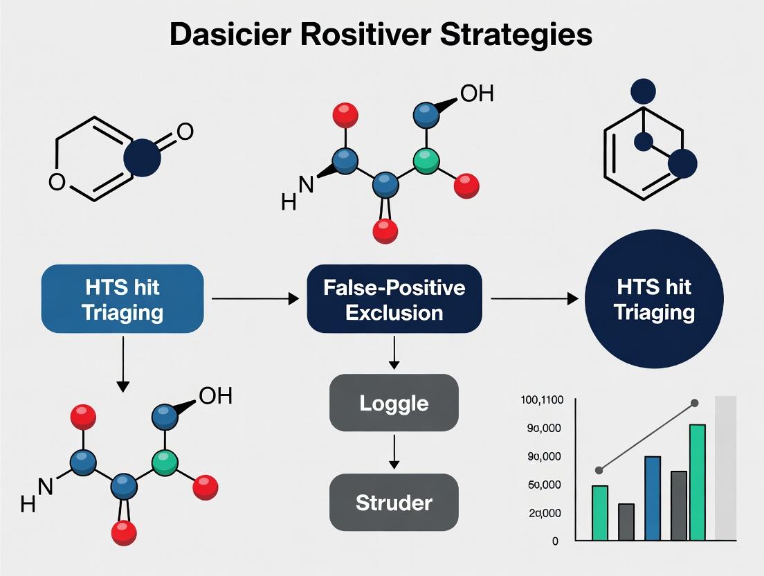
HTS Hit Triage: A Modern Guide to False-Positive Exclusion and Hit Validation in Drug Discovery
This article provides a comprehensive guide for researchers and drug development professionals on triaging hits from High-Throughput Screening (HTS) campaigns.
HTS Hit Triage: A Modern Guide to False-Positive Exclusion and Hit Validation in Drug Discovery
Abstract
This article provides a comprehensive guide for researchers and drug development professionals on triaging hits from High-Throughput Screening (HTS) campaigns. It details systematic strategies to identify and exclude false positives stemming from assay interference, compound aggregation, and promiscuous inhibition. The scope covers foundational knowledge of false-positive mechanisms, a step-by-step methodological framework for triage, advanced troubleshooting for complex cases, and rigorous validation techniques to prioritize genuine lead candidates. By integrating current best practices, this guide aims to improve the efficiency and success rate of early-stage drug discovery.
Understanding the HTS Hit Triage Landscape: Why False Positives Arise and How to Spot Them
In high-throughput screening (HTS), hit triage is the critical process of distinguishing true, promising chemical starting points from false positives and non-viable leads. The stakes are immense: poor triage can waste months of research and millions of dollars on compounds that ultimately fail. This technical support center addresses common experimental challenges within the thesis context of developing robust triaging strategies to systematically exclude false positives.
FAQs & Troubleshooting Guides
Q1: Our HTS campaign yielded a high hit rate (>5%). How do we prioritize which hits to triage first to avoid resource waste? A1: Implement a multi-parameter prioritization score. Common causes of high hit rates include promiscuous aggregators, compound interference, and non-specific cytotoxicity. Immediate steps:
- Calculate Ligand Efficiency (LE) and Lipophilic Ligand Efficiency (LLE): Prioritize hits with LE > 0.3 and LLE > 5. They offer better optimization potential.
- Check for structural alerts: Use tools like PAINS (Pan-Assay Interference Compounds) filters to flag problematic chemotypes.
- Review primary assay artifacts: Correlate hit potency with assay signal-to-noise (S/N) and Z'-factor from the primary screen. Hits from plates with Z' < 0.5 should be deprioritized.
Q2: We observe a loss of potency in dose-response confirmation compared to the single-concentration primary screen. What are the likely causes? A2: This is a classic sign of false positives.
- Cause 1: Compound precipitation or aggregation at higher concentrations. This causes non-specific inhibition.
- Troubleshooting: Perform a dynamic light scattering (DLS) assay. Re-test suspect hits with the addition of 0.01% Triton X-100; true targets are inhibited, while aggregator effects are reduced.
- Cause 2: Assay interference (e.g., fluorescence, quenching, reactivity).
- Troubleshooting: Run the assay in the presence of the compound but without the target/biologic component. A significant signal change indicates direct compound interference.
Q3: Hits confirm in biochemical assays but fail in cell-based counter-screens. What's the next step? A3: This suggests a lack of cellular permeability, engagement, or off-target cytotoxicity.
- Step 1: Perform a cytotoxicity assay (e.g., CellTiter-Glo) in parallel. Exclude hits with a cytotoxicity IC50 within 10-fold of the on-target activity IC50.
- Step 2: Assess membrane permeability via computational logP/logD predictions or experimental PAMPA assays. Hits with predicted logD > 4 or poor PAMPA permeability may have delivery issues.
- Step 3: Design a target engagement probe assay (e.g., CETSA, cellular thermal shift assay) to confirm the compound binds the intended target in cells.
Experimental Protocols for Key Triage Experiments
Protocol 1: Aggregator Detection Using DLS and Detergent Challenge Objective: Identify compounds that act via non-specific aggregation. Materials: See "Research Reagent Solutions" table. Method:
- Prepare a 10x stock of the hit compound in 100% DMSO. Dilute into assay buffer to a final concentration 10x above its reported Ki/IC50. Use a final DMSO concentration of ≤1%.
- Incubate at room temperature for 30 minutes.
- Transfer 100 µL to a low-volume quartz cuvette or DLS plate.
- Measure particle size distribution using a DLS instrument. Scan from 0.3 nm to 10,000 nm.
- Particles with a hydrodynamic radius > 100 nm suggest aggregation.
- Detergent Challenge: Repeat the primary assay with and without 0.01% v/v Triton X-100. A right-shift in IC50 > 10-fold with detergent confirms an aggregator mechanism.
Protocol 2: Counter-Screen for Fluorescence Interference (Fluorescence-Based Assays) Objective: Rule out compounds that modulate assay signal by affecting fluorescence. Method:
- In a black assay plate, prepare serial dilutions of the hit compound in assay buffer without the enzyme/substrate.
- Add the fluorescent probe/tracer at the concentration used in the primary assay.
- Incubate under primary assay conditions (time, temperature).
- Read the plate using the same excitation/emission settings as the primary screen.
- A concentration-dependent change in fluorescence signal indicates direct compound interference. Correct the primary assay data using these values or exclude the hit.
Data Presentation
Table 1: Common HTS Hit Triaging Counterscreens and Outcomes
| Triaging Assay | Purpose | Typical Positive Result Indicating False Positive | Acceptance Criteria for Progression |
|---|---|---|---|
| Dose-Response Confirmation | Verify concentration-dependent activity | No sigmoidal curve, IC50 not within 3x of primary screen value | Robust Hill slope (~1), IC50 confirmed, R² > 0.9 |
| DLS / Detergent Challenge | Detect non-specific aggregators | Particles >100 nm, IC50 shift >10-fold with detergent | No large particles, activity resistant to detergent |
| Fluorescence Interference | Detect signal artifacts | Change in fluorescence in target-less system | Signal change < ±10% of control |
| Cytotoxicity (Cell-Based) | Exclude general cell killers | Cytotoxicity IC50 < 10x on-target IC50 | Cytotoxicity IC50 > 10x on-target IC50 or no toxicity |
| Orthogonal Assay (e.g., SPR) | Confirm direct binding | No binding sensorgram, KD > 10 µM | Measurable binding, KD < 10 µM |
| PAINS Filter | Identify problematic chemotypes | Match to known PAINS substructures | No substructure matches |
Diagrams
Diagram 1: Core HTS Hit Triage Workflow
Diagram 2: Key Mechanisms of HTS False Positives
The Scientist's Toolkit: Research Reagent Solutions
Table 2: Essential Reagents for Hit Triage Experiments
| Reagent/Material | Function in Triage | Key Consideration |
|---|---|---|
| Triton X-100 | Non-ionic detergent used in aggregator challenge assays. Disrupts compound aggregates. | Use at low concentration (0.01%) to avoid target denaturation. |
| DMSO (Ultra-Pure, Hyroscopic) | Universal compound solvent. Must be high purity to prevent artifacts. | Keep water-free; final assay concentration typically 0.1-1%. |
| CellTiter-Glo / MTS Reagents | Luminescent/colorimetric assays for quantifying cell viability/cytotoxicity. | Choose based on assay compatibility (lytic vs. non-lytic). |
| SPR Chips (e.g., CM5, NTA) | Sensor surfaces for biophysical binding confirmation (Surface Plasmon Resonance). | Chip type must match target properties (e.g., NTA for His-tagged proteins). |
| PAMPA Plate System | Predictive parallel artificial membrane permeability assay for passive permeability. | Use brain or lipid-specific membranes for CNS programs. |
| Reference Controls (Known Inhibitor, Aggregator) | Critical for assay validation and comparator analysis (e.g., for DLS, detergent challenge). | Include in every triage experiment plate. |
Troubleshooting Guides & FAQs
FAQ 1: My high-concentration compound shows strong inhibition, but activity is lost upon dilution. What is the most likely cause, and how do I confirm it? Answer: This is a classic sign of compound aggregation. Aggregators non-specifically inhibit enzymes by forming colloidal particles that sequester proteins.
- Confirmatory Protocol (Dynamic Light Scattering):
- Prepare a 10-50 µM solution of your test compound in assay buffer (ensure DMSO concentration is ≤1%).
- Filter the solution using a 0.22 µm centrifugal filter to remove dust.
- Load the filtrate into a clean DLS cuvette.
- Measure the particle size distribution at 25°C. Run triplicate measurements.
- Interpretation: A population of particles in the 50-1000 nm range confirms aggregation. Compare to a known non-aggregator control.
FAQ 2: My compound's activity is inconsistent across different assay readouts (e.g., fluorescence vs. luminescence). What should I suspect? Answer: This strongly suggests assay interference, specifically optical interference (inner filter effect, fluorescence quenching/emission) or chemical interference (reactivity with assay components).
- Confirmatory Protocol (Readout Interference Counter-Screen):
- Fluorescence Interference: In the absence of the enzyme/target, mix the compound at its assay concentration with the fluorogenic substrate. Measure signal vs. substrate-only control. A significant signal shift indicates interference.
- Luminescence Interference: Perform the same null-target test with luciferin and ATP. Compare signal to controls.
- General Detergent Test: Repeat your primary assay in the presence of 0.01-0.1% v/v Triton X-100 or CHAPS. True inhibitors retain activity; aggregators often see inhibition abolished.
FAQ 3: My hit inhibits multiple, structurally unrelated targets in the panel. Is this a valuable pan-inhibitor or a false positive? Answer: It is likely a promiscuous inhibitor. This behavior can stem from aggregation, reactivity, or membrane disruption, and is generally undesirable for a selective lead.
- Confirmatory Protocol (Covalent/Reactive Promiscuity Check - Gel Shift Assay):
- Incubate a model protein (e.g., 5 µM serum albumin or lysozyme) with 50-100 µM test compound in PBS for 2-4 hours at 25°C.
- Include a control with DMSO only.
- Run samples on a non-reducing SDS-PAGE gel.
- Stain with Coomassie Blue.
- Interpretation: A higher molecular weight shift for the compound-treated sample suggests covalent modification/protein adduct formation.
FAQ 4: How can I quickly prioritize hits to deprioritize assay interference artifacts early? Answer: Implement a standardized orthogonal assay cascade. Hits from a primary biochemical assay must be confirmed in a secondary, biophysical or cell-based assay with a different readout technology.
Table 1: Prevalence of False-Positive Mechanisms in HTS Campaigns
| Mechanism | Typical Prevalence in HTS (%) | Key Characteristic | Common Diagnostic |
|---|---|---|---|
| Aggregation | 15-30% | Steep dose-response, Hill slope >1.5 | DLS, detergent sensitivity |
| Assay Interference | 5-20% | Readout-dependent activity, unstable time-course | Counter-screens, orthogonal assays |
| Promiscuous Inhibition | 5-15% (of actives) | Inhibits >3 unrelated targets | Panel screening, gel shift, redox cycling tests |
| Chemical Reactivity | 2-10% | Reacts with nucleophiles (Cys, Lys) | NMR/LC-MS adduct detection, thiol scavenger assay |
Table 2: Key Experimental Conditions to Modulate for Mechanism Identification
| Suspect Mechanism | Variable to Test | Expected Result for True Inhibitor | Expected Result for False Positive |
|---|---|---|---|
| Aggregation | Add 0.01% Triton X-100 | Activity maintained | Inhibition abolished or significantly reduced |
| Aggregation | Increase enzyme concentration 10-fold | IC50 shifts modestly | IC50 shifts dramatically (potency loss) |
| Redox/Chelation | Add 1-10 mM DTT or EDTA | Activity maintained | Inhibition abolished (redox cycler/chelator) |
| Covalent Binding | Pre-incubate, then dilute | Activity maintained (if reversible) | Inhibition persists (if irreversible) |
Experimental Protocols
Protocol: Detergent-Based Counter-Screen for Aggregation Objective: To distinguish specific inhibitors from aggregate-based inhibitors. Materials: Assay buffer, test compound(s), DMSO, 10% Triton X-100 stock, substrate, enzyme, detection reagents. Procedure:
- Prepare a 2X solution of test compound in assay buffer containing either 0.02% Triton X-100 (final 0.01%) or no detergent.
- In a 96-well plate, add 25 µL of the 2X compound solution (with or without detergent) to respective wells.
- Initiate the reaction by adding 25 µL of a 2X enzyme/substrate mixture.
- Run the assay according to standard primary conditions.
- Analysis: Plot dose-response curves with and without detergent. A rightward shift of >3-fold in IC50 with detergent suggests aggregate-based inhibition.
Protocol: Thiol-Reactivity Fluorescent Probe Assay Objective: Detect compounds that act as non-specific electrophiles. Materials: Test compound, 10 mM NAC (N-acetyl cysteine) in PBS, 100 µM mBBr (monobromobimane) in DMSO, PBS buffer, black 384-well plate. Procedure:
- In a low-volume plate, mix 10 µL of 100 µM compound with 10 µL of 500 µM NAC (final [NAC] = 250 µM). Include NAC-only and compound-only controls.
- Incubate for 1 hour at room temperature.
- Add 5 µL of 300 µM mBBr (final [mBBr] = 50 µM). mBBr fluoresces upon reacting with free thiols.
- Incubate for 15 min protected from light.
- Measure fluorescence (λex/λem ~380/460 nm).
- Analysis: A decrease in fluorescence in the compound+NAC sample vs. the NAC-only control indicates the compound consumed NAC via thiol reactivity.
The Scientist's Toolkit: Key Research Reagent Solutions
| Item | Function & Application |
|---|---|
| Triton X-100 / CHAPS | Non-ionic detergents used to disrupt compound aggregates in biochemical assays. A key tool for diagnosing aggregation. |
| DTT (Dithiothreitol) | Reducing agent. Used to identify redox-cycling compounds or inhibitors whose activity is reliant on oxidizing assay conditions. |
| N-Acetyl Cysteine (NAC) | Thiol-containing nucleophile. Serves as a mimic for protein cysteine residues to test for non-specific electrophilic reactivity. |
| BSA (Bovine Serum Albumin) | Inert protein. Added to assays to quench promiscuous inhibitors that act via colloidal aggregation or non-specific adsorption. |
| α-Lytic Protease | A sensitive, well-characterized enzyme often used in promiscuity panels. Inhibition of this model protease suggests non-specific mechanisms. |
| Dynamic Light Scattering (DLS) Plate Reader | Instrument for measuring hydrodynamic radius of particles in solution. Gold standard for directly confirming compound aggregation. |
Diagrams
HTS Hit Triage and False-Positive Exclusion Workflow
Classification of Assay Interference Mechanisms
Mechanism of Inhibition by Compound Aggregation
Technical Support Center: Troubleshooting False Positives in HTS
Troubleshooting Guides & FAQs
Q1: Our primary HTS shows high hit rates (>5%). What are the most common causes and initial checks? A1: A high hit rate is a primary red flag. Immediate checks should include:
- Assay Interference: Test compounds for fluorescence, absorbance, or quenching at assay wavelengths.
- Compound Integrity: Check for compound precipitation or aggregation using light scattering.
- Positive Control Normalization: Verify that your positive control (e.g., 100% inhibition) remains consistent across all plates.
- Reagent Stability: Ensure key reagents (enzymes, substrates) have not degraded.
Protocol 1: Aggregation-Induced Artifact Check
- Method: Perform a detergent-based rescue experiment.
- Steps:
- Run the primary assay in parallel with and without a non-ionic detergent (e.g., 0.01% Triton X-100 or Tween-20).
- For putative hits, re-test in a dose-response with and without detergent.
- Interpretation: A significant rightward shift (weakening) of the dose-response curve in the presence of detergent suggests the activity is caused by colloidal aggregation, a classic false-positive mechanism.
Q2: We observe a "steep" Hill slope (nH > 2.5) in dose-response curves. Does this indicate a problem? A2: Yes. Non-physiological Hill slopes often indicate assay interference or non-specific binding mechanisms, not true target engagement.
Protocol 2: Mechanistic Counter-Screen for Promiscuous Inhibitors
- Method: Use a orthogonal, non-enzymatic assay format.
- Steps:
- Test all hits with steep Hill slopes in a secondary, biophysical assay (e.g., Surface Plasmon Resonance - SPR, or Thermal Shift Assay - TSA).
- For enzymatic assays, re-test using an alternative substrate or a different detection technology (switch from fluorescence to luminescence).
- Interpretation: Lack of activity in the orthogonal assay confirms the hit is likely a false positive specific to the primary assay's detection method.
Q3: Hit activity is inconsistent between single-point and dose-response confirmations. How should we proceed? A3: This indicates poor assay robustness or compound instability.
- Action: Re-prepare compounds from dry powder for confirmation. Implement stringent plate quality controls: calculate Z'-factor for each screening plate. A Z' < 0.5 indicates an unreliable assay. Also, check for edge effects or evaporation by visualizing plate heat maps of raw signal.
Q4: What are key chemical structure alerts for false positives? A4: Certain chemotypes are notorious for non-specific activity. Flag these for priority counter-screening:
| Chemical Alert | Potential Mechanism | Recommended Counter-Screen |
|---|---|---|
| Pan-Assay Interference Compounds (PAINS) | Redox-activity, covalent modification, aggregation | Use a PAINS filter database; assay in presence of DTT; perform reactivity assay. |
| Compounds with Michael Acceptors | Covalent, non-specific binding | Assay in presence of excess nucleophile (e.g., β-mercaptoethanol). |
| Chelators (e.g., catechols) | Metal ion depletion | Add excess required metal cofactor (e.g., Mg²⁺, Mn²⁺). |
| Highly conjugated/fluorescent compounds | Optical interference | Test for signal in the absence of biological target. |
| Reactive esters/halides | Acylation/alkylation | Incubate with target in denatured state; check for time-dependent inhibition. |
The Scientist's Toolkit: Key Research Reagent Solutions
| Item | Function in False-Positive Triage |
|---|---|
| Non-ionic Detergent (Triton X-100) | Disrupts colloidal aggregates, confirming or denying aggregation-based inhibition. |
| Dithiothreitol (DTT) | Reducing agent; quenches signals from redox-active compounds (common PAINS). |
| Bovine Serum Albumin (BSA) or Lysozyme | Non-target protein; tests for non-specific protein binding or sequestration. |
| Control Enzyme (e.g., AmpC β-lactamase) | "Off-target" enzyme assay; identifies promiscuous enzyme inhibitors. |
| Alternative Substrate (Orthogonal Chemistry) | Rules out interference with the specific substrate or reporter used in the primary screen. |
| SPR Chip with Immobilized Target | Directly measures binding affinity and kinetics, independent of functional assay artifacts. |
Experimental Workflows & Pathway Diagrams
Title: HTS Hit Triage Workflow for False-Positive Exclusion
Title: Mechanism of Colloidal Aggregate Interference
Technical Support Center
This support center provides guidance for researchers navigating false-positive signals in High-Throughput Screening (HTS). Efficient troubleshooting is critical to mitigate the severe impact of false positives on project timelines, budgets, and the integrity of lead qualification.
Troubleshooting Guides & FAQs
Q1: Our HTS campaign yielded an unusually high hit rate (>5%). What are the first steps to triage for assay interference? A: A high hit rate is a primary indicator of potential systematic false positives. Implement this immediate triage protocol:
- Confirmatory Re-Test: Re-test the primary hits in the same assay format. False positives often are not reproducible.
- Counter-Screen: Immediately run a orthogonal assay measuring a different readout but related biology. Hits active in both are more likely true.
- Artifact Check: For fluorescence/luminescence-based assays, test compounds at hit concentrations for intrinsic fluorescence, quenching, or luciferase inhibition.
- Protocol: Intrinsic Fluorescence Check
- Prepare hit compounds at the screening concentration in assay buffer.
- Measure fluorescence/luminescence in the absence of all assay reagents using the same plate reader settings.
- A signal >3 standard deviations above buffer-alone control indicates potential interference.
Q2: How can we quickly distinguish aggregator false positives from true bioactive compounds? A: Compound aggregation is a major source of false-positive inhibition. Perform these diagnostic experiments:
- Protocol: Detergent Sensitivity Test
- Re-run the enzymatic or binding assay for a subset of hits under two conditions: Standard buffer vs. Buffer containing 0.01%-0.1% Triton X-100 or Tween-20.
- Interpretation: If inhibitory activity is abolished or significantly reduced in the presence of detergent, it strongly suggests aggregation-based inhibition.
- Protocol: Dynamic Light Scattering (DLS)
- Prepare hit compounds at 10-50 µM in assay buffer.
- Analyze using a DLS instrument. The presence of particles in the 100-1000 nm range confirms aggregation.
Q3: Our cell-based assay hits are cytotoxic, confounding the target-specific signal. How do we deconvolute this? A: Cytotoxicity is a frequent confounder in phenotypic screens. A multi-parametric approach is required.
- Viability Counter-Screen: Run a parallel cell viability assay (e.g., ATP-based luminescence, resazurin reduction) under identical seeding and dosing conditions.
- Time-Dependence: Measure the primary assay readout at multiple earlier time points (e.g., 6h, 12h, 24h). Cytotoxic effects often manifest later than target-specific modulation.
- Cytotoxicity Index Table: Use data to calculate an index for prioritization.
| Hit ID | Primary Assay IC₅₀ (µM) | Viability Assay CC₅₀ (µM) | Selectivity Index (CC₅₀/IC₅₀) | Triage Action |
|---|---|---|---|---|
| FP-101 | 1.2 | 1.5 | 1.25 | Exclude. Activity likely secondary to cell death. |
| LQ-202 | 0.5 | >20 | >40 | Priority. Specific effect confirmed. |
| A-303 | 2.0 | 8.0 | 4.0 | Caution. Requires further mechanistic studies. |
Q4: What orthogonal assays are most definitive for false-positive exclusion before lead declaration? A: A cascade of orthogonal tests is the gold standard.
- Biophysical Confirmation: Use Surface Plasmon Resonance (SPR) or Isothermal Titration Calorimetry (ITC) to confirm direct, stoichiometric binding to the purified target.
- Genetic Corroboration: In cell assays, use CRISPR knockout or RNAi knockdown of the target. True hits will show diminished activity in genetically modified cells.
- Chemical Response: Assess activity against known target mutants (for kinases, etc.) or with competitive ligands to confirm expected mechanism.
Experimental Protocols for Key Triage Experiments
Protocol: Orthogonal Assay Cascade for Hit Validation Objective: To sequentially exclude false positives using independent methodologies. Workflow:
- Input: Primary HTS Hit List.
- Step 1 - Dose-Response in Primary Assay: Confirm potency and curve shape. Reject irreproducible or flat curves.
- Step 2 - Artifact/Interference Assays: Perform detergent, fluorescence, and cytotoxicity counter-screens (see protocols above). Reject artifacts.
- Step 3 - Secondary Orthogonal Assay: Use a different readout/technology (e.g., switch from FP to TR-FRET, or biochemical to phenotypic). Reject inactive compounds.
- Step 4 - Biophysical Binding: Validate direct binding via SPR. Reject non-binders.
- Output: Orthogonally Verified Lead Series.
Title: Hit Triage Workflow for False Positive Exclusion
Protocol: Target Engagement Assay in Cells (CETSA Principle) Objective: Provide evidence of target binding in a physiologically relevant cellular environment. Method:
- Treat intact cells with compound or DMSO control.
- Heat challenge cells at a specific temperature (e.g., 55°C) to denature proteins.
- Lyse cells and separate soluble (native) from insoluble (denatured) protein via centrifugation.
- Quantify target protein in the soluble fraction by immunoblotting or AlphaLisa.
- Interpretation: True binders stabilize the target, increasing its amount in the soluble fraction post-heating compared to DMSO control.
Title: Cellular Target Engagement (CETSA) Workflow
The Scientist's Toolkit: Essential Research Reagent Solutions
| Item | Function & Application in False-Positive Triage |
|---|---|
| Non-Ionic Detergents (Triton X-100, Tween-20) | Diagnose aggregator false positives by disrupting colloidal aggregates in biochemical assays. |
| AlphaLisa/AlphaScreen Beads | Enable homogeneous, no-wash assays for secondary orthogonal screening; reduce interference from colored/fluorescent compounds. |
| Cellular Viability Assay Kits (e.g., CellTiter-Glo) | Essential for parallel cytotoxicity counter-screening in cell-based HTS. |
| SPR Biosensor Chips (e.g., CM5, NTA) | Provide label-free, quantitative confirmation of direct compound-target binding kinetics (KD, kon, koff). |
| Tagged Recombinant Protein (His-tag, GST-tag) | Required for immobilization in biophysical binding assays (SPR, ITC) and secondary biochemical assays. |
| CRISPR sgRNA Libraries/Knockout Cell Lines | Genetically validate target specificity; loss of compound activity in KO cells confirms on-target mechanism. |
| Thermal Shift Dyes (e.g., SYPRO Orange) | Used in Differential Scanning Fluorimetry (DSF) to assess compound-induced protein stabilization, a sign of binding. |
Technical Support & Troubleshooting Center
Frequently Asked Questions (FAQs)
Q1: When querying PubChem BioAssay, I receive too many hits from phenotypic assays that don't specify a molecular target. How can I filter for target-based HTS data relevant to my protein of interest?
A: Use the 'Target Name' filter with specific gene symbols or UniProt IDs. Combine this with the 'Assay Type' filter set to 'Confirmatory' or 'Screening' and 'Target Type' set to 'Protein'. For example, to find assays for kinase MAPK1, use the Advanced Search query: "MAPK1"[Target Name] AND "confirmatory"[Assay Type]. This will exclude many broad phenotypic screens.
Q2: How do I handle conflicting bioactivity values (e.g., Ki vs. IC50) for the same compound-target pair across ChEMBL and PubChem? A: Prioritize data based on assay confidence. Follow this decision workflow:
- Prefer values from direct binding assays (e.g., Ki from radioligand binding) over functional assays (IC50).
- Within functional assays, prefer orthogonal assay types (e.g., fluorescence + luminescence) confirming the same activity.
- Check the 'Data Validity Comment' field in ChEMBL and 'Activity Outcomes/Issues' in PubChem for flags like 'Inconclusive' or 'Non-specific inhibitor'.
- Use the consensus value or report the range. A structured approach is in Table 2.
Q3: My compound shows high potency in a primary HTS but is absent from major databases. How can I perform an initial literature-based liability assessment? A: Conduct a systematic search using the compound's SMILES or InChIKey in:
- PubMed: Search
"[InChIKey]" AND (toxicity OR reactive OR pan-assay OR PAINS). - Google Scholar: Search the structural scaffold or core name with terms like "frequent hitter" "assay interference" "cytotoxicity".
- Specialized Resources: Manually check the Non-Obvious Molecule Archive (NOMA) and SureChEMBL for patent-derived activity and potential reactive groups.
Troubleshooting Guides
Issue: Inconsistent SAR when merging datasets from PubChem BioAssay AID 404 and ChEMBL. Symptoms: A compound series shows a clear potency trend in one database but appears scattered or inactive in the other. Diagnosis & Resolution:
- Verify Assay Protocols: Extract and compare the experimental parameters. Critical differences often include:
- Assay readout (e.g., fluorescence intensity vs. time-resolved FRET).
- Target construct (e.g., full-length protein vs. catalytic domain).
- Cofactor/compound pre-incubation time.
- Check Concentration Ranges: The active range in one assay may be outside the tested range in the other.
- Normalize Data: Convert all activity values to pActivity (-log10 of molar concentration). Apply a standardized cutoff (e.g., pActivity > 5 for actives). See Protocol 1 for dataset merging.
Issue: High rate of promiscuous (frequent-hitter) compounds passing initial HTS triage from PubChem data. Symptoms: Hits are active across multiple disparate target families in PubChem BioAssay, suggesting non-specific mechanisms. Diagnosis & Resolution:
- Cross-Database Profiling: Query the hit structure in ChEMBL's 'Compound Report Card' to view the Target Heatmap. Genuine selective compounds typically show activity against closely related targets.
- Apply Computational Filters: Before purchasing/resynthesizing, screen SMILES against:
- PAINS filters: Using the RDKit or CDK implementation.
- Aggregation predictors: Use the
Aggregator Advisortool (available via the University of Kansas website). - Covalent/Reactive Group Checkers: Use SMARTS patterns from literature (e.g., Baell & Holloway, 2010).
- Validate Experimentally: Employ a counter-screen for aggregation (e.g., detergent addition assay; see Protocol 2).
Data Presentation
Table 1: Core Database Comparison for HTS Triaging
| Feature | PubChem BioAssay | ChEMBL |
|---|---|---|
| Primary Focus | Repository of individual HTS assays, including primary & summary results. | Curated bioactivity data extracted from literature, focused on drug discovery. |
| Data Type | Raw assay data, dose-response curves, protocol details. | Standardized activities (Ki, IC50), target assignments, derived SAR. |
| Key Triaging Utility | Access to primary HTS readouts to assess assay interference. | Target-centric view of compound promiscuity and lead-likeness metrics. |
| False-Positive Flagging | Yes, via "Activity Outcomes/Issues" tags (e.g., "inconclusive"). | Yes, via "Data Validity Comment" (e.g., "Non-specific inhibitor"). |
| Linkage to Compounds | Direct link to PubChem Compound for properties. | Integrated with drug metabolism and ADMET data where available. |
Table 2: Resolving Conflicting Bioactivity Data
| Scenario | Priority (1=Highest) | Recommended Action for Thesis Triaging |
|---|---|---|
| Ki (binding) vs. IC50 (functional) both reported. | 1. Ki | Use Ki for binding affinity claims. Note functional potency (IC50) separately. |
| IC50 values differ >10-fold between sources. | 2. Orthogonal Assays | Prefer value from the assay with a non-optical readout (e.g., SPR, radiometric). |
| Only discrepant IC50 values exist. | 3. Most Recent & Curated | Prefer ChEMBL's curated value over a single PubChem entry. |
| One source flags as "inconclusive". | 4. Unflagged Data | Discard the flagged datum; proceed with the unflagged value with caution. |
Experimental Protocols
Protocol 1: Merging & Profiling Hit Data from PubChem and ChEMBL
Objective: Create a unified dataset for SAR analysis while tagging data provenance.
- Data Retrieval: For your hit list, download bioactivity data via:
- PubChem: Use the
PUG-RESTAPI with theassay/activityendpoint. - ChEMBL: Use the
chembl_webresource_clientin Python or the web interface 'Target Search'.
- PubChem: Use the
- Standardization: Convert all activity values to pChEMBL/pActivity scale:
pActivity = -log10(activity_value_in_molar). - Merging: Use the
InChIKeyas the primary key to merge tables. Add columns forsource_databaseandassay_type. - Flagging: Add a
conflict_flagcolumn. Tag entries where pActivity differs by >1.5 units (≈30-fold difference) for the same compound-target pair. - Output: A unified CSV file with columns:
InChIKey,SMILES,Target,pActivity,Activity_Type,Source_DB,Assay_Description,Conflict_Flag.
Protocol 2: Detergent-Based Counter-Screen for Aggregation-Based False Positives
Objective: Determine if compound activity is due to colloidal aggregation.
- Materials: Hit compound(s), positive control aggregator (e.g., tetracycline), assay buffer, non-ionic detergent (Triton X-100 or Nonidet P-40).
- Procedure: a. Run the primary HTS assay protocol in duplicate. b. Include a condition with 0.01% v/v final concentration of Triton X-100 in the assay buffer. c. Test compounds at their previously determined IC50/Ki concentration.
- Analysis: A significant reduction (>50%) in activity in the detergent condition strongly suggests the activity is aggregation-mediated. The compound should be deprioritized.
- Note: Include detergent-only controls to ensure it does not interfere with the assay signal.
Mandatory Visualization
Diagram 1: HTS Hit Triaging & False-Positive Exclusion Workflow
The Scientist's Toolkit: Research Reagent Solutions
| Item | Function in HTS Triaging Context |
|---|---|
| Non-ionic Detergent (Triton X-100/Nonidet P-40) | Used in aggregation counter-screens to disrupt colloidal aggregates, identifying compound-based assay interference. |
| Redox-Sensitive Dye (e.g., DTT, TCEP) | Quenches reactive oxygen species; used to test if compound activity is due to redox-cycling, a common false-positive mechanism. |
| Albumin (BSA or HSA) | Added to assay buffer to test for compound sequestration by plasma proteins, an early ADMET liability check. |
| Fluorescent Probe (Reference Ligand) | Used in orthogonal displacement assays (e.g., FP, TR-FRET) to confirm target binding in a different detection modality. |
| CYP450 Isozyme Kits | For early metabolic stability screening of triaged hits to prioritize compounds with a higher chance of in vivo stability. |
| Cytotoxicity Assay Kit (e.g., MTT, CellTiter-Glo) | Essential to rule out that activity in a cell-based assay is due to general cell death rather than specific target modulation. |
A Step-by-Step Triage Protocol: From Primary Hits to Confirmed Actives
Troubleshooting Guides & FAQs
Q1: Our High-Throughput Screening (HTS) identified a hit compound with strong signal in the primary assay, but it shows high absorbance/fluorescence at the assay wavelength. What is the first step to confirm if this is an optical interference false positive?
A1: Perform a straightforward control experiment: a full wavelength scan of the compound at your assay concentration in the buffer used. Compare the scan to the emission/excitation spectra of your assay's detection method (e.g., fluorophore). Overlap indicates potential interference. Next, run a mock assay (all components except the biological target). A persistent signal confirms optical interference.
Experimental Protocol: Wavelength Scan & Mock Assay
- Prepare the compound at the same concentration used in the HTS in the assay buffer.
- Using a plate reader or spectrophotometer, perform an absorbance scan from 200 nm to 700 nm.
- For fluorescence, perform an emission scan at the HTS excitation wavelength and an excitation scan at the HTS emission wavelength.
- Prepare assay plates with all reagents (buffer, substrate, co-factors, detection reagents) excluding the enzyme or cellular target.
- Add the hit compound and controls (DMSO, known inhibitor).
- Run the assay using the HTS protocol and measure signal.
Q2: After ruling out optical interference, how do I determine if the compound is a promiscuous aggregator, a common source of false positives?
A2: Utilize two complementary assays: 1) The detergent sensitivity test, and 2) Dynamic Light Scattering (DLS). Aggregators often inhibit enzymes non-specifically by forming particles that sequester proteins.
Experimental Protocol: Detergent Sensitivity Test
- Perform a dose-response of the hit compound in your primary biochemical assay.
- Run the identical dose-response in parallel with the addition of a non-ionic detergent (e.g., 0.01% Triton X-100 or 0.01% Tween-20).
- A significant rightward shift (weakening) of inhibition in the presence of detergent is a strong indicator of aggregation-based inhibition.
Experimental Protocol: Dynamic Light Scattering (DLS)
- Prepare the hit compound at 10-50x its IC50 concentration in the assay buffer.
- Incubate for the same duration as the assay.
- Measure particle size distribution using a DLS instrument. The presence of particles in the 100-1000 nm range, absent in buffer-only controls, confirms aggregation.
Q3: The hit passes initial interference and aggregator tests but is inactive in a cell-based counter-screen. Could it be a compound reactivity (PAINS) issue or a permeability problem?
A3: This requires a bifurcated approach to differentiate between chemical reactivity and lack of cellular penetration.
A. Testing for Reactivity (Redox/Antioxidant Activity):
- Experimental Protocol: DTT-Based Redox Assay: Incubate the compound with 1 mM DTT (dithiothreitol) in buffer for 1-2 hours. Analyze by LC-MS for adduct formation or decomposition. Reactivity with this nucleophilic thiol suggests potential pan-assay interference.
- Experimental Protocol: Glutathione (GSH) Trapping Assay: Incubate the compound with liver microsomes/NADPH and glutathione. Detect and quantify glutathione adducts using LC-MS/MS. This identifies electrophilic, potentially reactive metabolites.
B. Testing for Cellular Permeability:
- Experimental Protocol: Parallel Artificial Membrane Permeability Assay (PAMPA): Use a commercial PAMPA plate to measure passive diffusion. Low permeability suggests the compound may not effectively enter cells.
- Critical Control: Ensure the cell-based assay uses a relevant, engineered cell line expressing the target of interest, confirmed via qPCR or immunoblotting.
Data Presentation
Table 1: Summary of Primary Triage Assays for False-Positive Exclusion
| Triage Parameter | Assay Name | Key Measurement | Positive Result Indicative of False Positive | Typical Follow-Up |
|---|---|---|---|---|
| Optical Interference | Full Wavelength Scan | Absorbance/Fluorescence Spectrum | Overlap with assay detection wavelengths | Reformulate assay or use label-free technology. |
| Optical Interference | Mock Assay (No Target) | Signal Output | Signal above background | Disqualify or seek orthogonal assay format. |
| Compound Aggregation | Detergent Sensitivity Test | IC50 Shift with Detergent | >10-fold shift in IC50 | Characterize further with DLS; consider solubility improvements. |
| Compound Aggregation | Dynamic Light Scattering (DLS) | Hydrodynamic Diameter | Particles >100 nm | Disqualify or investigate aggregator-prone chemotypes. |
| Chemical Reactivity | DTT/GSH Adduct Assay | Adduct formation (LC-MS) | Covalent modification of compound | Classify as potential PAINS; prioritize other chemotypes. |
| Cellular Activity | Cell-Based Counter-Screen | Efficacy (e.g., Viability, Reporter) | No activity at >10x biochemical IC50 | Investigate permeability (PAMPA), efflux, or compound instability. |
| Target Specificity | Orthogonal Biophysical Assay (SPR, ITC) | Binding Affinity (KD) | No binding observed | Disqualify; primary assay artifact confirmed. |
Table 2: Key Research Reagent Solutions for Hit Triage
| Item | Function in Triage | Example/Supplier |
|---|---|---|
| Non-Ionic Detergents (Triton X-100, Tween-20) | Disrupts compound aggregates; key reagent for aggregator identification. | Sigma-Aldrich, Thermo Fisher |
| Dithiothreitol (DTT) | Reducing agent used to test for redox-cycling or thiol-reactive compounds. | Gold Biotechnology, Cayman Chemical |
| Reduced Glutathione (GSH) | Nucleophilic tripeptide for trapping reactive electrophilic metabolites. | MilliporeSigma, BioVision |
| PAMPA Plate System | Measures passive permeability of compounds through an artificial lipid membrane. | Corning Gentest, pION |
| SPR Biosensor Chips (e.g., CM5) | Surface for immobilizing target protein to measure direct binding kinetics via Surface Plasmon Resonance. | Cytiva |
| Label-Free Detection Reagents (e.g., MST-capillaries, ITC cells) | Enable binding assays without fluorescent/radioactive labels, avoiding optical interference. | NanoTemper, Malvern Panalytical |
Mandatory Visualizations
Title: Optical Interference Triage Pathway
Title: Aggregator & Reactivity Triage Pathway
Title: Sequential Multi-Parameter Triage Funnel Workflow
Troubleshooting Guides & FAQs
Q1: Why is my primary HTS hit showing activity in a counter-screen against an unrelated target? A: This is a classic sign of a promiscuous, aggregation-based false positive. These compounds form colloidal aggregates that non-specifically inhibit a wide range of enzymes. To troubleshoot, repeat the assay in the presence of a non-ionic detergent (e.g., 0.01% Triton X-100 or 0.1 mg/mL CHAPS). A significant reduction or loss of activity confirms aggregation.
Q2: My compound passes the detergent test but still shows high hit rates in orthogonal assays with different readouts. What could be the cause? A: The compound may be interfering with the assay technology itself (assay artifact). Common culprits include:
- Fluorescence/ Luminescence Interference: The compound may be a quencher, absorber, or auto-fluorescent at the assay wavelengths.
- Redox Cyclers: The compound may be generating reactive oxygen species that inactivate the target or assay reagents.
- Chelators: It may be sequestering essential metal co-factors. Troubleshoot by:
- Running an interference assay with the signal-generating system in the absence of the biological target.
- Using a label-free, biophysical method (e.g., SPR, ITC) to confirm direct binding.
Q3: What is the recommended follow-up if a compound shows steep, non-linear dose-response curves in the primary screen? A: Steep curves (Hill coefficient >> |1|) often indicate a mechanism not related to specific, reversible target inhibition. This can signal denaturation, precipitation, or redox activity. Immediately prioritize the following orthogonal assays:
- Cytotoxicity Assay: Rule out general cell death as the mechanism in cell-based screens.
- Thiol Reactivity Assay: Test against a panel of cysteine-containing proteins (e.g., glutathione, albumin) to identify pan-assay interference compounds (PAINS).
- LC-MS Analysis: Verify compound integrity under assay conditions to rule out degradation.
Q4: How do I differentiate true target engagement from off-target effects in a cellular phenotype screen? A: This requires a multi-tiered approach. First, confirm the phenotype is concentration-dependent and reproducible. Then, employ:
- Chemical/Genetic Perturbation: Use a known tool compound or siRNA against your target. If the phenotype is not mimicked or rescued, your hit is likely acting off-target.
- Cellular Thermal Shift Assay (CETSA): This directly measures target engagement in a cellular lysate or live cells, providing evidence the compound binds the intended protein.
- Photoaffinity Labeling/Proteomics: For completely novel targets, this can help identify the actual protein(s) the compound is binding to.
Q5: My orthogonal assay contradicts my primary HTS result. Which result should I trust? A: Generally, trust the result from the more direct, biophysical, and label-free method. Assays with fewer components and simpler readouts (e.g., NMR, SPR, DSF) are less prone to artifacts than complex, amplified biochemical or phenotypic assays. The primary HTS result should be considered a hypothesis until confirmed by an orthogonal method with a different detection principle.
Key Quantitative Data for Hit Triage
Table 1: Typical Rates and Characteristics of Common False Positives
| False Positive Class | Approximate Frequency in HTS* | Key Characteristic | Primary Orthogonal Assay |
|---|---|---|---|
| Aggregators | 5-20% | Loss of activity with detergent | Re-test with 0.01% Triton X-100 |
| Fluorescent/Luminescent Interferers | 2-10% | Signal in target-free control | Counter-screen with signal-only system |
| Redox Cyclers/Reactive Compounds | 1-5% | Steep Hill slope, time-dependent | DTT/GSH reactivity assay; LC-MS stability |
| PAINS (Pan-Assay Interference Compounds) | 3-8% | Hits in multiple, unrelated assays | Structural filters; thiol reactivity assays |
| Cytotoxic Compounds | 1-15% (cell-based) | Correlation with viability readouts | Viability assay (e.g., ATP content) |
*Frequency is library and assay-dependent. These are generalized estimates.
Table 2: Comparison of Orthogonal Assay Technologies
| Assay Type | Principle | Throughput | Cost | Key Application in Triage |
|---|---|---|---|---|
| Surface Plasmon Resonance (SPR) | Direct binding measurement | Medium | High | Confirm direct target binding; measure kinetics |
| Differential Scanning Fluorimetry (DSF) | Thermal stabilization of target | Medium-High | Low | Confirm binding; assess ligand stability |
| Cellular Thermal Shift Assay (CETSA) | Target engagement in cells | Medium | Medium | Confirm cellular target engagement |
| Interference Assay | Signal measurement sans target | High | Very Low | Rule out technology-specific artifacts |
| Liquid Chromatography-Mass Spec (LC-MS) | Compound integrity analysis | Low | Medium-High | Confirm compound stability in assay buffer |
Experimental Protocols
Protocol 1: Aggregation Counter-Screen using Detergent
Objective: To determine if inhibitory activity is due to colloidal aggregate formation. Materials: Hit compound(s), DMSO, assay buffer, non-ionic detergent (e.g., Triton X-100), primary assay reagents. Method:
- Prepare two identical sets of serial dilutions of the hit compound in DMSO.
- Dilute the first set into standard assay buffer. Dilute the second set into assay buffer containing a final concentration of 0.01% (v/v) Triton X-100.
- Run the primary HTS assay protocol with both sets of compound solutions in parallel.
- Analysis: Calculate IC50 values for both conditions. A right-shift of >3-fold (increase in IC50) or complete loss of potency in the detergent condition strongly suggests the compound acts via aggregation.
Protocol 2: Fluorescence Interference Assay
Objective: To identify compounds that interfere with fluorescence or luminescence readouts. Materials: Hit compound(s), DMSO, assay plate, fluorogenic/luminogenic substrate (without enzyme/target), appropriate buffer, plate reader. Method:
- In a black or white assay plate, dilute compounds to the same top concentration used in the primary HTS. Include DMSO-only controls.
- Add the signal-generation reagent (substrate) in buffer to all wells. Do not add the enzymatic target.
- Incubate under primary assay conditions (time, temperature).
- Read the plate using the same instrument settings (ex/em wavelengths, gain) as the primary HTS.
- Analysis: A signal change >20% of the DMSO control signal indicates the compound is interfering with the detection technology.
Protocol 3: Liquid Chromatography-Mass Spectrometry (LC-MS) for Compound Integrity
Objective: To verify the hit compound is stable under assay conditions. Materials: Hit compound, DMSO, assay buffer, LC-MS system. Method:
- Prepare the hit compound at 10x its assay concentration in DMSO.
- Dilute this stock 1:10 into pre-warmed assay buffer (final DMSO = 1%). Also prepare a control in buffer only.
- Incubate the mixture at the assay temperature (e.g., 25°C or 37°C) for the duration of the assay incubation time.
- At time = 0 and time = incubation end, quench the reaction (e.g., by adding equal volume of cold acetonitrile). Centrifuge to pellet precipitated protein/buffer salts.
- Inject the supernatant into the LC-MS. Analyze the chromatogram and mass spectra for the parent compound peak.
- Analysis: A significant decrease in the parent peak area or the appearance of new major peaks indicates compound degradation.
The Scientist's Toolkit: Research Reagent Solutions
| Item | Function in False-Positive Triage |
|---|---|
| Non-Ionic Detergents (Triton X-100, CHAPS) | Disrupts colloidal aggregates formed by promiscuous inhibitors, used to confirm/rule out aggregation-based mechanisms. |
| Dithiothreitol (DTT) / Glutathione (GSH) | Reducing agents used to test if a compound's activity is due to redox cycling or reactivity with thiol groups. |
| Bovine Serum Albumin (BSA) or Human Serum Albumin (HSA) | Used to assess non-specific binding; significant potency shift in the presence of protein suggests compound promiscuity. |
| Fluorogenic/Luminogenic Substrate (Target-Free) | The core reagent for running interference assays to identify technology-specific false positives. |
| Cytotoxicity Assay Kit (e.g., ATP-based) | Essential for cell-based screens to decouple specific target modulation from general cell death. |
| Stable, Purified Target Protein | Required for all biophysical confirmation assays (SPR, DSF, ITC) to prove direct binding. |
Diagrams
DOT Script for Hit Triage Workflow
Title: Rapid False-Positive Triage Workflow
DOT Script for PAINS & Reactivity Pathways
Title: Common Mechanisms of PAINS and Reactive Compounds
Troubleshooting Guides & FAQs
Q1: During LC-MS analysis for hit validation, I observe a mass corresponding to my compound of interest, but also a significant +22 Da adduct peak. What is this, and how does it impact triaging?
A: The +22 Da peak is typically a sodium adduct [M+Na]+. This is common in electrospray ionization (ESI) when sodium ions are present in the solvent or sample. While it confirms the molecular weight, its presence at high intensity can suppress the protonated [M+H]+ ion, leading to inaccurate quantification of the actual compound's abundance. For triaging, ensure your calibration standards and samples use the same solvent system (e.g., avoid sodium-containing buffers for MS). Use ammonium formate/acetate buffers instead to promote [M+H]+ or [M+NH4]+ formation. Re-analyze with cleaned sample preparation to exclude false positives from salt aggregates.
Q2: My 1H NMR spectrum of a triaged HTS hit shows extra singlets and broad peaks in the aromatic region, but LC-MS shows >95% purity. What could be the issue?
A: LC-MS at >95% purity suggests the issue is not a major contaminant. The extra peaks likely indicate:
- Residual Solvent or Water: Check DMSO-d6/H2O peaks. Dry the sample thoroughly.
- Degradation: The compound may degrade in the NMR tube. Prepare the sample in deuterated solvent and acquire spectra immediately. Compare with LC-MS data from the same solution over time.
- Conformers/Atropisomers: Broad peaks suggest slow interconversion on the NMR timescale. Try variable-temperature NMR. For triaging, this signals potential instability, which is a critical exclusion criterion. Proceed to stability assays.
Q3: In UHPLC-UV purity analysis, what is an acceptable threshold for a single unidentified impurity when prioritizing hits for further study?
A: For early-stage triaging of HTS hits, the following thresholds are commonly applied to exclude false positives from compound degradation or synthesis errors:
| Analysis Method | Typical Acceptable Threshold for a Single Unidentified Impurity | Rationale for Hit Triaging |
|---|---|---|
| UHPLC-UV (214-254 nm) | ≤ 2.0% | Impurities >2% can complicate SAR, suggest instability, or be responsible for the bioactivity. |
| LC-MS (TIC, UV 254 nm) | ≤ 5.0% (with no single impurity >2%) | Higher tolerance as MS is more specific, but major impurities require identification. |
Q4: My compound shows a clean 1H NMR and good LC-MS purity, but biological activity is inconsistent. What orthogonal purity method should I use?
A: This is a classic sign of a highly potent minor impurity. Implement orthogonal methods:
- Quantitative NMR (qNMR): Uses an internal standard (e.g., dimethyl terephthalate) to absolutely quantify the active component versus all other protons.
- LC-MS with Evaporative Light Scattering Detection (ELSD) or Charged Aerosol Detection (CAD): These are mass-sensitive detectors that provide a response independent of chemical structure, unlike UV, revealing impurities with poor chromophores.
Experimental Protocol: Orthogonal Purity Assessment by qNMR
- Materials: Purified compound, deuterated solvent (e.g., DMSO-d6), qNMR standard (e.g., 99.9% maleic acid).
- Procedure:
- Precisely weigh 1-10 mg of your compound and ~1 mg of the qNMR standard into an NMR tube.
- Add 0.6 mL of deuterated solvent.
- Acquire a standard 1H NMR spectrum with sufficient relaxation delay (e.g., 25-30 seconds).
- Integrate a well-resolved, unique proton signal from your compound and a unique signal from the standard.
- Calculate purity: Purity (%) = (Icmpd / Ncmpd) / (Istd / Nstd) × (Wstd / Wcmpd) × Pstd × 100.
- I = Integral, N = Number of protons for the signal, W = Weight, Pstd = Purity of standard.
The Scientist's Toolkit: Key Research Reagent Solutions
| Item | Function in Integrity/Purity Analysis |
|---|---|
| LC-MS Grade Solvents (Acetonitrile, Methanol, Water) | Minimize background noise and ion suppression in MS, ensuring accurate molecular ion detection. |
| Deuterated NMR Solvents (DMSO-d6, CDCl3) | Provide a field frequency lock and allow for proper solvent signal suppression during NMR acquisition. |
| qNMR Standard (e.g., Maleic Acid) | High-purity internal standard for absolute quantitation of the target compound via proton NMR. |
| Ammonium Formate/Acetate (MS Grade) | Volatile buffers for LC-MS that promote clean [M+H]+/[M-H]- ionization and prevent sodium/potassium adducts. |
| Silica Gel/Prep TLC Plates | For rapid micro-scale purification of hits from DMSO stock to remove degradants before analysis. |
| 0.22 µm PTFE Syringe Filters | Essential for removing particulate matter from samples prior to LC-MS/UHPLC injection, protecting the column. |
Workflow & Relationship Diagrams
Hit Integrity & Purity Assessment Workflow
Analytical Signatures of Common False Positive Causes
Troubleshooting Guide & FAQs
Q1: In SPR, my sensogram shows a high response in the reference flow cell, leading to poor double-referenced data. What could be the cause? A: This is often due to non-specific binding of the analyte to the sensor chip surface or the dextran matrix. To troubleshoot:
- Increase buffer ionic strength: Use 150-300 mM NaCl in the running buffer.
- Add a non-ionic detergent: Include 0.005% (v/v) P20 surfactant in buffers.
- Use a different chip chemistry: Switch to a carboxylated (CM) series chip and optimize immobilization pH to reduce positive charge, or use a hydrophobic capture (HPA) chip if non-specific binding is to lipids.
- Include a blocking step: After ligand immobilization, inject a 1-3 minute pulse of 1 M ethanolamine-HCl (pH 8.5) to block unreacted groups.
Q2: My ITC experiment shows very low heat change (ΔH), making data analysis unreliable. How can I improve the signal? A: Low heat change typically indicates weak binding affinity or low concentration.
- Optimize concentrations: The product c = Ka * [M]total * n should be between 10 and 500 for reliable fitting. For weak binders (Ka < 10^4 M⁻¹), use high macromolecule concentration (e.g., 100-500 µM).
- Check buffer matching: Ensure the ligand and macromolecule are in EXACTLY the same buffer (pH, salt, DMSO%). Dialyze the macromolecule against the buffer, then use the dialysis buffer to prepare the ligand solution.
- Increase cell temperature: Performing the experiment at 25°C or 37°C instead of 15°C can increase the enthalpic signal for some interactions.
Q3: In CETSA, I see no thermal stabilization of the target protein even with a high concentration of a confirmed binder. What might be wrong? A: Lack of stabilization can be due to assay conditions that do not reflect cellular context or compound behavior.
- Verify cellular permeability: Ensure your compound is cell-permeable. Use a known cell-permeable positive control (e.g., a clinical inhibitor of your target).
- Check incubation time & temperature: Insufficient time for compound engagement can cause this. Extend compound incubation (e.g., 1-2 hours at 37°C) before heating.
- Confirm target abundance: Ensure your detection method (Western blot, MS) is sensitive enough for the endogenous protein levels. Overexpression systems may be needed for low-abundance targets.
- Optimize heating temperature: The chosen heating temperature must cause partial denaturation/unfolding of the target. Perform a full melt curve (e.g., 37°C to 67°C) to find the appropriate T_m.
Experimental Protocols
Protocol 1: Surface Plasmon Resonance (SPR) – Capture Coupling for Proteins
- Chip Preparation: Use a Series S CMS chip. Prime the system with filtered, degassed HBS-EP+ buffer (10 mM HEPES, 150 mM NaCl, 3 mM EDTA, 0.05% v/v Surfactant P20, pH 7.4).
- Ligand Immobilization: Dilute anti-GST antibody in 10 mM sodium acetate (pH 4.5) to 20 µg/mL. Inject for 60 seconds at 10 µL/min to achieve ~10,000 RU capture level. Inject your GST-tagged protein at 5-10 µg/mL in running buffer for 120-300 seconds to achieve optimal ligand density (50-100 RU for kinetics).
- Analyte Binding: Perform 2-fold serial dilutions of the analyte in running buffer (+ matching DMSO%). Inject each concentration for 120s (association) followed by 300s dissociation (flow rate: 30 µL/min). Include a zero-concentration blank for double referencing.
- Regeneration: Inject 10 mM glycine-HCl (pH 1.5) for 30-60 seconds to regenerate the surface without damaging the captured antibody.
Protocol 2: Isothermal Titration Calorimetry (ITC) – Protein-Small Molecule Interaction
- Sample Preparation: Dialyze the protein into ITC buffer (e.g., PBS, pH 7.4) overnight. Use the final dialysis buffer to prepare the compound solution, ensuring perfect buffer matching. Centrifuge both samples (14,000 x g, 10 min) before loading to remove particulates.
- Instrument Setup: Degas both samples for 10 minutes. Fill the cell with protein (typical concentration 10-100 µM). Load the syringe with compound (typical concentration 10-20x higher than the cell). Set temperature to 25°C, reference power to 10 µCal/s, and stirring speed to 750 rpm.
- Titration Program: Perform an initial 0.4 µL injection (discarded in analysis), followed by 19 injections of 2.0 µL each, with 150-second spacing between injections.
- Data Analysis: Integrate raw heat peaks, subtract the dilution heat from the compound into buffer, and fit the binding isotherm to a one-site binding model to derive Ka, ΔH, ΔS, and n (stoichiometry).
Protocol 3: Cellular Thermal Shift Assay (CETSA) – Western Blot Endpoint Format
- Compound Treatment: Seed cells in 10-cm dishes. Treat with compound or DMSO vehicle for desired time (e.g., 2 hours).
- Heated Fraction Preparation: Harvest cells by trypsinization, wash with PBS, and resuspend in PBS with protease inhibitors. Aliquot 100 µL of cell suspension into PCR tubes.
- Heat Challenge: Heat aliquots at defined temperatures (e.g., 42, 47, 52, 57, 62°C) for 3 minutes in a thermal cycler, followed by 3 minutes at 25°C.
- Lysis & Analysis: Lyse cells by three freeze-thaw cycles in liquid nitrogen. Centrifuge (20,000 x g, 20 min, 4°C). Collect soluble fraction and analyze by SDS-PAGE and Western blotting for your target protein. Quantify band intensity to generate melting curves.
Table 1: Comparison of Target Engagement Biophysical Methods
| Parameter | SPR | ITC | CETSA |
|---|---|---|---|
| Measurement | Binding kinetics & affinity (kon, koff, K_D) | Thermodynamics (K_A, ΔG, ΔH, ΔS, n) | Thermal stability shift (ΔTm, apparent KD) |
| Sample Consumption | Low (µg of protein) | High (mg of protein) | Cell lysate or intact cells |
| Throughput | Medium-High | Low | Medium |
| Key Artifact Source | Non-specific binding, mass transport | Buffer mismatch, low heat signal | Compound permeability, protein aggregation |
| Context | Purified system | Purified system | Cellular environment |
| Typical K_D Range | pM - mM | nM - mM | nM - µM (cellular) |
Table 2: Common CETSA Buffers and Additives
| Reagent | Purpose/Function |
|---|---|
| PBS (for harvesting) | Maintains physiological pH and osmolarity during cell washing. |
| Protease Inhibitor Cocktail | Prevents target protein degradation during sample preparation and heating. |
| NP-40 (0.1-0.4%) | Mild detergent used in some lysis buffers to aid in soluble fraction recovery post-heating. |
| TCEP (1 mM) | Reducing agent to prevent protein aggregation via disulfide bonds during heating. |
| Cycloheximide (50 µg/mL) | Optional: added pre-treatment to inhibit new protein synthesis, focusing on pre-existing pool. |
The Scientist's Toolkit: Key Research Reagent Solutions
| Item | Function in TE Studies |
|---|---|
| Biacore Series S Sensor Chips (CM5, CAP) | Gold surface with carboxymethylated dextran for covalent ligand immobilization or capture. |
| Anti-GST Antibody | For capture-coupling of GST-tagged proteins on SPR chips, preserving protein activity. |
| MicroCal ITC Standard Cells | High-sensitivity calorimetry cells for measuring heat changes upon binding. |
| CETSA-Compatible Lysis Buffer | Buffer optimized to maintain protein solubility after thermal challenge for WB or MS readout. |
| Protease Inhibitor Cocktail (EDTA-free) | Essential for CETSA to prevent degradation, especially after heat-induced unfolding. |
| High-Affinity Positive Control Inhibitor | A well-characterized binder for your target to serve as a positive control in all TE assays. |
| DMSO (Molecular Biology Grade) | Universal compound solvent; must be matched in all samples and controls (<0.5-1% final). |
Visualization: Experimental Workflows and Pathways
Diagram 1: SPR Kinetic Analysis Workflow
Diagram 2: CETSA Principle & Signal Pathway
Diagram 3: Hit Triaging Strategy for False-Positive Exclusion
Troubleshooting Guides and FAQs for SAR and Analogue Testing
Q1: During SAR testing, we see a dramatic loss of activity in all newly synthesized analogues of the initial hit. What could be the cause? A: This often indicates the initial hit was a false-positive or that the core pharmacophore was incorrectly identified. First, re-test the original compound to confirm its activity. Then, verify the synthetic pathway and purity of your analogues via LC-MS. If purity is confirmed, the issue may be excessive structural modification. Return to the simplest possible analogue (e.g., a single atom substitution) to establish a baseline.
Q2: Our dose-response curves in the confirmatory assay are irreproducible and show high variability. How can we troubleshoot this? A: High variability often stems from compound handling or assay conditions.
- Compound Solubility: Ensure all analogues are in true solution. Use a standardized solvent (e.g., DMSO) and perform serial dilution in assay buffer, monitoring for precipitation. Use a consistent final DMSO concentration (typically ≤0.1-1%).
- Assay Artifacts: Re-run the assay including control wells with DMSO-only and a known inhibitor/activator. Check for edge effects on the plate. Ensure the cell passage number or enzyme batch is consistent.
- Data Analysis: Use a robust curve-fitting model (e.g., four-parameter logistic) and ensure sufficient data points across the concentration range.
Q3: How do we differentiate between true SAR trends and results caused by compound interference (e.g., assay interference, aggregation)? A: Implement a set of counter-screens and control experiments.
- For Optical Assays: Run a fluorescence or absorbance read of the compound alone at the test concentration.
- For Aggregation: Add non-ionic detergents (e.g., 0.01% Triton X-100) to the assay buffer; true inhibitors are typically detergent-insensitive.
- Orthogonal Assays: Confirm activity in a biophysical or functional assay with a different readout (e.g., SPR, thermal shift, cell-based reporter).
Q4: What is the recommended number of analogues to synthesize for early SAR? A: There is no fixed number, but a focused library of 10-20 compounds is typical for initial exploration. The goal is to test specific hypotheses about key structural features.
Table 1: Key Metrics for Early SAR Analysis
| Metric | Target Value | Purpose & Notes |
|---|---|---|
| Number of Analogues Synthesized | 10 - 20 | Balances exploration with resource allocation. |
| Purity Threshold (LC-MS) | ≥95% | Essential for interpreting biological data. |
| Confirmed Potency (IC50/EC50) | <10 µM (for a µM hit) | Establifies credible biological effect. |
| Selectivity Index (vs. related target) | >10-fold | Early indicator of specificity. |
| Ligand Efficiency (LE) | >0.3 kcal/mol per heavy atom | Assesses binding efficiency of the core structure. |
Detailed Experimental Protocol: SAR by Analogue Testing
Title: Protocol for Synthesis and Evaluation of a Focused Analogue Library
Objective: To systematically modify a high-throughput screening (HTS) hit and evaluate the impact on biological activity to establish preliminary SAR and exclude false positives.
Materials & Reagents:
- Parent HTS hit compound.
- Chemical reagents for synthesis (as per design).
- Analytical HPLC/MS system.
- Assay plates, buffers, and detection reagents from the original confirmatory HTS assay.
- Orthogonal assay kit/materials (e.g., SPR chip, thermal shift dye).
Procedure:
- Analogue Design: Design analogues to probe specific regions of the hit: (A) The core scaffold (ring modifications), (B) Linker regions (length, flexibility), (C) Peripheral substituents (steric, electronic effects).
- Chemical Synthesis: Synthesize target analogues using appropriate organic chemistry techniques. Record all reaction conditions and yields.
- Compound Characterization & Purity Analysis:
- Purify compounds using flash chromatography or HPLC.
- Analyze each analogue by LC-MS. Confirm molecular weight and achieve ≥95% purity.
- Prepare 10 mM stock solutions in DMSO. Store at -20°C.
- Biological Re-Testing:
- Primary Confirmatory Assay: Test all analogues in the same assay used to validate the original hit. Use an 8-point, 1:3 serial dilution (e.g., from 30 µM to 0.001 µM) in duplicate.
- Counter-Screen Assays: Run all active analogues (IC50/EC50 < 10 µM) in the interference assays noted in FAQ A3.
- Orthogonal Validation: Select the 2-3 most promising analogues for testing in a biophysical or secondary functional assay.
- Data Analysis:
- Calculate potency (IC50/EC50) and efficacy (% inhibition/activation) for each compound.
- Calculate ligand efficiency: LE = (-RT ln(IC50))/HA, where HA is the number of non-hydrogen atoms.
- Compile data into a table (see Table 1) and create SAR summary diagrams.
The Scientist's Toolkit: Key Research Reagent Solutions
Table 2: Essential Materials for SAR & Analogue Testing
| Item | Function in SAR | Example/Note |
|---|---|---|
| High-Purity DMSO | Universal solvent for compound stock solutions. | Use anhydrous, sterile DMSO. Aliquot to prevent water absorption. |
| LC-MS Grade Solvents | For compound purification and analysis. | Essential for accurate purity assessment. |
| Assay-Ready Plates | High-quality microplates for dose-response. | Low binding plates recommended for precious compounds. |
| Cryogenic Vials | Long-term storage of compound stocks. | Use for master stock archive at -80°C. |
| Non-Ionic Detergent (Triton X-100) | To test for compound aggregation. | Use at 0.01% final concentration in assay buffer. |
| Fluorescence Quencher (e.g., Trypan Blue) | To identify inner filter effects in fluorescence assays. | Quenches external fluorescence signal. |
| Standardized Control Compound | Reference for assay performance and data normalization. | Known potent agonist/antagonist for the target. |
| SPR Chip or Thermal Shift Dye | For orthogonal, biophysical confirmation of binding. | Select based on target properties (protein size, stability). |
Visualizations
Diagram 1: SAR & Analogue Testing Workflow for Hit Triaging
Diagram 2: Key SAR Testing Pathways & Decision Logic
Troubleshooting Guides & FAQs
Q1: Our dose-response curve has a poor fit (low R²), making IC50 determination unreliable. What are the common causes and solutions? A: Poor curve fitting often stems from:
- Insufficient Data Points: Use a minimum of 10 concentrations, spanning at least 5 orders of magnitude around the suspected IC50.
- Incorrect Model Selection: Ensure you are using the correct equation (e.g., 4-parameter logistic (4PL) for standard inhibition, 5PL for asymmetric curves). For enzyme kinetics, use the Morrison equation for tight-binding inhibitors to derive Ki.
- Outliers or Edge Effects: Check for evaporation in edge wells or compound precipitation at high concentrations. Use plate layouts that randomize treatments.
Q2: The calculated IC50 values vary significantly between replicate experiments. How can we improve reproducibility? A: High variability indicates assay instability or inconsistent protocol execution.
- Key Checks:
- Enzyme/Cell Stability: Ensure consistent passage number, thawing procedures, and incubation times.
- Compound Handling: Use fresh DMSO stocks, avoid freeze-thaw cycles, and confirm compound solubility in assay buffer.
- Signal Window: Maintain a robust Z'-factor (>0.5) for the assay plate. A shrinking signal window invalidates potency measurements.
Q3: How do we distinguish a true inhibitor from a non-specific aggregator or a fluorescent interferer at this stage? A: This is a critical triage step to exclude false positives.
- For Aggregators: Perform a detergent challenge (e.g., add 0.01% Triton X-100). A right-shift in IC50 (>3-fold) suggests aggregation-based inhibition.
- For Spectroscopic Interference: Run a control plate with substrate and inhibitor but no enzyme/cells to detect signal quenching or enhancement.
- Orthogonal Assay: Confirm activity using a biophysical method (e.g., SPR, ITC) or a functional assay with a different readout (e.g., cell-based vs. biochemical).
Q4: When should we use IC50, and when is Ki more appropriate? A: IC50 is an empirical, assay-dependent value. Ki, the inhibition constant, is a true molecular binding constant.
- Use IC50 for initial potency ranking under your specific assay conditions.
- Calculate Ki when you need a comparable, mechanism-based metric for lead optimization. For competitive inhibitors, use the Cheng-Prusoff equation (IC50 = Ki*(1+[S]/Km)). For non-competitive or tight-binding inhibitors, more complex models (Morrison equation) are required.
Q5: Our compound appears to show >100% inhibition or a "hook effect" at high doses. What does this mean? A: This is a red flag for assay artifact or complex mechanism.
- >100% Inhibition: Suggests interference with the detection method (e.g., fluorescence quenching, compound absorbance).
- Hook Effect (Inhibition decreases at high concentration): Can indicate compound precipitation, cytotoxicity in cell assays, or alternative binding modes. Re-test solubility and include relevant controls.
Experimental Protocols
Protocol 1: 10-Point Dose-Response Curve for IC50 Determination (Biochemical Assay)
- Compound Dilution: Prepare a 100X top concentration in DMSO. Perform 1:3 serial dilutions in DMSO to create 10 stock concentrations.
- Plate Preparation: Using an Echo or pintool, transfer 50 nL of each DMSO stock into a 384-well assay plate in triplicate. Include DMSO-only (0% inhibition) and control inhibitor (100% inhibition) wells.
- Reaction Addition: Add 5 µL of enzyme in assay buffer to all wells. Incubate for 10 minutes.
- Initiate Reaction: Add 5 µL of substrate (at concentration ≈ Km) to start the reaction.
- Readout: Monitor product formation kinetically (e.g., every minute for 30 min) using a plate reader (absorbance, fluorescence).
- Data Analysis: Calculate reaction velocity for each well. Fit normalized velocity vs. log[compound] data to a 4PL model: Y=Bottom + (Top-Bottom)/(1+10^((LogIC50-X)*HillSlope)).
Protocol 2: Determining Ki for a Tight-Binding Inhibitor Using the Morrison Equation
- Experimental Design: Run dose-response curves at multiple enzyme concentrations ([E]) around the suspected Ki (e.g., 0.5x, 1x, 2x, and 4x the estimated Ki).
- Assay Execution: Perform the enzymatic assay as in Protocol 1, ensuring initial velocity conditions.
- Global Fitting: Fit the combined dataset to the Morrison equation for tight-binding inhibition: vi/v0 = 1 - (([E]+[I]+Kiapp) - sqrt(([E]+[I]+Kiapp)^2 - 4[E][I]))/(2[E]), where Ki_app = Ki*(1+[S]/Km).
- Output: The fitting software will output the true Ki value, which should be consistent across the different [E] used.
Data Presentation
Table 1: Troubleshooting Common Dose-Response Curve Issues
| Symptom | Potential Cause | Diagnostic Test | Solution |
|---|---|---|---|
| Shallow Hill Slope | Partial inhibition, cooperativity, multiple targets | Test in orthogonal assay; check purity | Use 5PL fit; purify compound |
| Hill Slope >1.5 | Compound aggregation, cooperative binding | Detergent challenge; DLS measurement | Add detergent; optimize buffer |
| IC50 varies with [E] | Tight-binding or irreversible inhibition | Run Morrison analysis; time-dependence | Report Ki, not IC50 |
| Poor curve fit (Low R²) | Too few points, outlier, wrong model | Inspect raw data | Use ≥10 points; remove outlier; try 5PL |
| Plate-to-plate variability | Cell/enzyme prep inconsistency, edge effects | Monitor Z'-factor, randomize layout | Standardize protocols, use plate seals |
Table 2: Key Reagent Solutions for Robust Dose-Response Experiments
| Reagent/Material | Function & Critical Specification | Example Vendor/Product |
|---|---|---|
| DMSO (Anhydrous) | Universal compound solvent. Must be high purity, low water content to prevent hydrolysis. | Sigma-Aldrich, D8418 |
| Assay Buffer (e.g., PBS, HEPES) | Maintains pH and ionic strength. Must be compatible with enzyme and detection method. | Thermo Fisher, J61136.AP |
| Control Inhibitor (Potent) | Provides 100% inhibition reference for curve normalization and QC. | Tocris Bioscience (various) |
| Detection Reagent | Quantifies reaction product (e.g., luminescent substrate, fluorescent dye). | Promega, CellTiter-Glo |
| Low-Volume Dispenser | Accurately transfers nanoliter compound doses (critical for DMSO tolerance). | Labcyte Echo / Beckman Biomek |
| 384-Well Microplates | Assay vessel. Must have low binding and be compatible with reader. | Corning, 3574 (White, solid) |
| Plate Reader | Measures assay signal. Requires precision for kinetic reads. | BioTek Synergy H1 / BMG CLARIOstar |
Mandatory Visualizations
Title: Hit Triage Workflow from IC50 to Ki
Title: Dose-Response Curve Equation Parameters
Advanced Troubleshooting for Problematic Hits and Optimizing Your Triage Workflow
In High-Throughput Screening (HTS) hit triaging, "sticky" or promiscuous compounds are a major source of false positives. These molecules nonspecifically inhibit or modulate multiple, often unrelated, targets through mechanisms distinct from classical, reversible binding. This technical support center provides troubleshooting guides and FAQs to help researchers confirm and exclude these problematic compounds, ensuring robust hit progression in drug discovery pipelines.
Troubleshooting Guides & FAQs
Q1: My primary HTS assay shows strong inhibition, but the compound appears inactive in all secondary orthogonal assays. Is this a sign of a promiscuous compound?
A: Yes, this is a classic red flag. Promiscuous aggregators or redox cyclers often show activity only in specific assay formats (e.g., certain detection chemistries). The recommended confirmation strategy is a multi-pronged orthogonal approach:
- Re-test in a detergent-supplemented assay: Add non-ionic detergent (e.g., 0.01% Triton X-100) to your primary assay buffer. Genuine inhibitors will retain activity, while aggregators often lose activity as micelles are disrupted.
- Perform a time-dependent assay: Many promiscuous mechanisms (e.g., chemical reactivity) show time-dependent inhibition. Compare IC50 values measured after 5-minute vs. 60-minute pre-incubation with the target. A significant leftward shift suggests a non-equilibrium mechanism.
- Conduct a counter-screen: Use a well-characterized, unrelated enzyme (e.g., β-lactamase) under identical buffer conditions. Inhibition of this control enzyme suggests nonspecific behavior.
Q2: How can I distinguish between a true hit and a compound that acts through redox cycling or fluorescence interference?
A: These are common assay artifacts. Follow this protocol:
Protocol: Confirmation of Redox Activity & Optical Interference
- Coupled Enzyme Redox Assay: In a 96-well plate, mix 50 µM compound, 100 µM NADH, and 50 µM WST-1 or MTT tetrazolium dye in phosphate buffer (pH 7.4). Monitor absorbance at 440 nm (WST-1) or 570 nm (MTT) for 30 minutes. A rapid increase indicates redox cycling activity.
- Fluorescence Quenching/Enhancement Test: Prepare assay plates with all components except the target enzyme. Scan the plate at your assay's excitation/emission wavelengths. A signal shift >10% of the DMSO control suggests direct optical interference.
- LC-MS/MS Validation: Post-assay, use LC-MS/MS to quantify the test compound and the expected product. The lack of expected product formation, despite compound depletion, can indicate it is being consumed in a side reaction.
Q3: What is the most definitive method to confirm target-specific binding for a compound suspected of being "sticky"?
A: Biophysical methods are considered the gold standard. Surface Plasmon Resonance (SPR) or Microscale Thermophoresis (MST) can confirm direct, stoichiometric binding.
Protocol: Confirmation via Surface Plasmon Resonance (SPR)
- Immobilization: Immobilize the purified target protein on a CM5 chip via amine coupling to achieve ~5000-10,000 Response Units (RU).
- Running Buffer: Use HBS-EP+ buffer (10 mM HEPES, 150 mM NaCl, 3 mM EDTA, 0.05% v/v Surfactant P20, pH 7.4). The surfactant reduces nonspecific binding.
- Kinetic Analysis: Inject a 3-fold dilution series of the compound (e.g., from 100 µM to 0.14 µM) at a flow rate of 30 µL/min. Include a solvent correction cycle.
- Data Interpretation: Analyze sensograms. A genuine binder will show concentration-dependent, rapid-on/rapid-off binding kinetics that fit a 1:1 binding model. Promiscuous compounds often show poorly fitting curves, massive carry-over, or non-equilibrium binding.
Data Presentation
Table 1: Summary of Promiscuity Mechanisms and Confirmatory Assays
| Mechanism | Primary Assay Signal | Key Confirmatory Assay | Expected Outcome for True Negative |
|---|---|---|---|
| Aggregation | Inhibition | Assay + 0.01% Triton X-100 | >10x increase in IC50 |
| Chemical Reactivity | Time-dependent Inhibition | Cysteine addition (1-10 mM) | Activity abolished |
| Redox Cycling | False activity in redox assays | NADH/WST-1 counter-screen | Rapid signal generation |
| Fluorescence Interference | False inhibition/activation | Fluorescence scan without enzyme | >10% signal change vs control |
| Membrane Disruption | Inhibition in cell-based assays | Lactate Dehydrogenase (LDH) release assay | Increased LDH release |
Table 2: Performance Metrics of Biophysical Confirmation Methods
| Method | Sample Consumption (Target) | Throughput | Key Readout for Promiscuity |
|---|---|---|---|
| SPR | ~50 µg (immobilization) | Medium | Nonsensical kinetics, poor wash-off |
| MST | ~5 µg (in solution) | Medium-High | Non-sigmoidal binding curve |
| DSF | <1 µg (in solution) | High | Non-specific protein destabilization |
| NMR (1H-15N HSQC) | >100 µg (in solution) | Low | Widespread chemical shift perturbations |
Experimental Protocols
Detailed Protocol: Detergent-Based Resistance Assay for Aggregators
- Prepare 2x assay buffer with and without 0.02% Triton X-100.
- Serially dilute the compound in 100% DMSO.
- In a 384-well plate, add 5 µL of compound dilution to wells (final DMSO = 0.5%).
- Add 20 µL of 2x assay buffer ± detergent to the appropriate wells.
- Pre-incubate compound with buffer for 15 minutes at RT.
- Initiate the reaction by adding 25 µL of 2x substrate/enzyme mixture (in standard buffer).
- Run the assay as normal and plot dose-response curves. Compare IC50 values.
Detailed Protocol: Differential Scanning Fluorimetry (DSF) for Nonspecific Binding
- Prepare a master mix containing purified target protein (2 µM) and SYPRO Orange dye (5X final) in assay buffer.
- Dispense 18 µL of master mix into each well of a 96-well PCR plate.
- Add 2 µL of test compound (in DMSO) or DMSO control for a final concentration of 50 µM compound.
- Seal the plate and centrifuge briefly.
- Run in a real-time PCR instrument: ramp temperature from 25°C to 95°C at a rate of 1°C/min, monitoring fluorescence.
- Calculate the melting temperature (Tm) shift. A large, nonspecific shift (>2°C) suggests compound "stickiness."
Mandatory Visualizations
Title: Hit Confirmation Workflow for Promiscuity Exclusion
Title: Mechanisms of Promiscuous Compound Action
The Scientist's Toolkit
Table 3: Key Research Reagent Solutions for Confirmation Experiments
| Reagent/Kit | Function in Confirmation | Key Consideration |
|---|---|---|
| Non-ionic Detergents (Triton X-100, Tween-20) | Disrupts compound aggregates, confirming aggregation-based inhibition. | Use at low concentration (0.01-0.1%) to avoid protein denaturation. |
| Redox-Sensitive Dyes (WST-1, MTT, Resazurin) | Detects redox cycling compounds by measuring reduction in a target-independent manner. | Run in the absence of target enzyme for a clean counter-screen. |
| β-Lactamase Enzyme | Serves as a well-characterized, generic enzyme for promiscuity counter-screens. | Choose a version not related to your target's protein family. |
| Surfactant P20 (for SPR) | Included in SPR running buffer to minimize nonspecific compound binding to the chip dextran. | Critical for analyzing "sticky" compounds; reduces false-positive sensorgrams. |
| SYPRO Orange Dye | Used in DSF to monitor protein unfolding; large Tm shifts indicate nonspecific compound binding. | Ensure the dye does not interact with the test compound itself. |
| LC-MS/MS Systems | Quantifies compound integrity and expected product formation post-assay, identifying compounds consumed in side reactions. | Requires authentic standards for both parent compound and expected product. |
Troubleshooting Guides & FAQs
Q1: During a high-throughput screen (HTS), my hit compound shows strong inhibition, but activity is lost upon re-testing. Could this be compound aggregation, and what is the first diagnostic test? A1: Yes, this is a classic symptom of a colloidal aggregation false positive. The first and most rapid diagnostic test is the Nonionic Detergent Challenge. Re-run the activity assay in the presence of a nonionic detergent like Triton X-100 or Tween-20 (typically at 0.01-0.02% v/v). Genuine inhibitors will retain activity, while aggregate-based inhibition is often abolished as the detergent disrupts the colloidal particles.
Q2: My compound fails the detergent test, suggesting aggregation. How do I confirm this with Dynamic Light Scattering (DLS) and what are the key parameters to interpret? A2: DLS measures particle size distribution in solution.
- Protocol: Prepare the compound at the assay concentration (typically 10-100 µM) in your assay buffer. Filter the buffer (0.1 µm filter) and compound stock solution to remove dust. Load sample into a cuvette, equilibrate at assay temperature, and run measurements (typically 10-15 sub-runs).
- Interpretation: A positive DLS signal for aggregation shows a major population of particles with hydrodynamic radii (Rh) > 50-100 nm. Monomeric small molecules are too small (< 2 nm) for DLS to detect reliably. Compare the intensity-weighted distribution to the volume- or number-weighted distributions; aggregates dominate the intensity signal.
Q3: What are the critical thresholds for DLS to confirm an aggregating compound? A3: See the table below for key quantitative indicators.
| Parameter | Typical Monomeric Compound | Indicative of Aggregation |
|---|---|---|
| Hydrodynamic Radius (Rh) | < 2 nm (often undetectable) | Primary peak > 50-100 nm |
| Polydispersity Index (PDI) | N/A or High (due to low signal) | Variable, often > 0.3 |
| Signal Intensity | Very Low | Significantly higher than buffer control |
| Effect of 0.01% Triton X-100 | No change | Major reduction in intensity/size of aggregate peak |
Q4: The detergent test is inconclusive for my compound. Are there other experimental mitigations to confirm or rule out aggregation? A4: Yes, employ a multi-pronged approach:
- Enzyme Concentration Dependence: Test inhibition at varying enzyme/target concentrations. Aggregator inhibition often decreases as target concentration increases, due to stoichiometric sequestration of the target on the aggregate surface.
- Time-Dependent Inhibition: Use pre-incubation experiments. True aggregators often show time-dependent inhibition as aggregates form.
- Alternative Detergents: Test with different detergents (e.g., CHAPS, NP-40) at critical micelle concentration (CMC).
- Ultrasound & Filtration: Briefly sonicate the compound solution or pass it through a low-binding 0.1 µm filter. If activity is lost, it suggests large, disruptive aggregates were responsible.
Q5: Within my thesis on HTS triage, at which stage should I implement these aggregation diagnostic assays? A5: Aggregation diagnostics should be integrated early in the post-HTS triage funnel, immediately after initial hit confirmation (see workflow diagram below). This prevents wasted resources pursuing artifactual hits.
Q6: What are the essential reagents and materials for setting up these diagnostics? A6: The Scientist's Toolkit:
| Research Reagent Solution | Function in Aggregation Diagnostics |
|---|---|
| Triton X-100 | Nonionic detergent; disrupts hydrophobic interactions in colloidal aggregates. |
| Tween-20 | Alternative nonionic detergent, sometimes less disruptive to some protein targets. |
| CHAPS | Zwitterionic detergent; useful for membrane-associated targets. |
| DLS Instrument | Measures hydrodynamic size distribution of particles in solution (e.g., Malvern Zetasizer). |
| Low-Binding Filters (0.1 µm) | Removes pre-formed aggregates from stock solutions for control experiments. |
| Ultrapure, Filtered Buffer | Essential for DLS to minimize background noise from dust particles. |
Experimental Protocols
Protocol 1: Nonionic Detergent Challenge Assay
- Prepare a 2X detergent solution in your assay buffer (e.g., 0.02% Triton X-100).
- In one set of assay wells, mix equal volumes of 2X compound and 2X detergent solution. In the control set, mix compound with buffer alone.
- Initiate the assay as normal by adding target/protein and substrate.
- Compare dose-response curves (IC50) with and without detergent. A right-shift of >10-fold is a strong indicator of aggregation.
Protocol 2: Dynamic Light Scattering (DLS) Measurement
- Buffer Preparation: Filter assay buffer through a 0.02 µm syringe filter into a clean glass vial. This is the background control.
- Sample Preparation: Dilute compound stock (DMSO) into filtered buffer to final assay concentration. Ensure final DMSO is consistent (< 1-2%).
- Measurement: Load 50-80 µL into a clean, low-volume cuvette. Place in instrument, equilibrate at 25°C for 2 minutes.
- Settings: Set number of sub-runs to 12, duration per sub-run automatically defined. Use "automatic attenuation" selection.
- Analysis: Measure buffer control first, then samples. Use instrument software to analyze intensity-, volume-, and number-weighted size distributions. Subtract the buffer control profile.
Diagrams
Title: HTS Triage Funnel with Aggregation Screening
Title: How Detergents Disrupt Aggregate-Based Inhibition
FAQs & Troubleshooting Guides
Q1: How can I differentiate a true hit from a compound acting as a redox cycler in my assay?
A: Redox cyclers generate reactive oxygen species (ROS), leading to signal drift and false positives in assays measuring fluorescence, luminescence, or cell viability. To triage them:
- Add Redox-Sensitive Dyes: Include cell-permeable dyes like CellROX (for ROS) in the assay. A dose-dependent increase in dye signal alongside your primary readout suggests redox activity.
- Conduct a Reducing Agent Challenge: Re-run the assay in the presence of a non-specific reducing agent like DTT (1-5 mM) or TCEP. True hits will be unaffected, while redox cycler signals will be quenched.
- Perform an Assay under Anaerobic Conditions: If the signal diminishes in an anaerobic chamber, it indicates oxygen-dependent redox cycling.
Q2: My fluorescent assay shows unexpected signal quenching. How do I determine if my compound is a fluorescent quencher?
A: Fluorescent quenchers (e.g., certain aromatic compounds, heavy atoms) interfere with the fluorophore, causing false negatives or distorted dose-responses.
- Direct Fluorophore Interaction Test: In a buffer-only plate, incubate your compound with the assay's fluorophore at its working concentration. Measure fluorescence. A decrease confirms direct quenching.
- Time-Resolved Fluorescence (TRF) or FP Assay Switch: Quenchers often affect intensity-based assays. Re-test hits in a time-resolved fluorescence (TRF) or fluorescence polarization (FP) format, which are less prone to these artifacts.
- Excitation/Emission Scan: Use a plate reader to scan the excitation/emission spectra of the fluorophore with and without the compound. A spectral shift or broad reduction indicates interference.
Q3: Suspected metal chelators are confounding my metalloenzyme assay. How do I confirm and exclude them?
A: Metal chelators deplete essential cofactors (e.g., Zn²⁺, Mg²⁺, Mn²⁺), non-specifically inhibiting many targets.
- Add-Back Experiment: Perform the assay with suspected hits. In parallel wells, add an excess of the relevant metal ion (e.g., 100 µM ZnSO₄ for zinc-dependent enzymes). Restoration of activity confirms chelation.
- Colorimetric Chelation Assay: Use a metal-sensitive dye like Zincon (for Zn²⁺) or Pyrocatechol Violet in a buffer. Compound-induced color change indicates chelation potential.
- Control with EDTA: Include EDTA as a control inhibitor. Compounds with a similar potency and mechanism (reversed by add-back) are likely chelators.
Q4: What is a consolidated experimental workflow for triaging these assay artifacts early in HTS?
A: Implement a counter-screen cascade immediately following primary HTS.
Title: HTS Hit Triaging Workflow for Artifact Exclusion
Table 1: Common Artifact Compounds and Their Diagnostic Signatures
| Artifact Class | Example Compounds | Diagnostic Test | Expected Artifact Signal | Reference IC50 Shift with Countermeasure |
|---|---|---|---|---|
| Redox Cyclers | Menadione, Paraquat, 5Z-7-Oxozeaenol | 5 mM DTT Challenge | >70% signal reduction | ≥10-fold right-shift |
| Metal Chelators | EGCG, TPEN, 8-Hydroxyquinoline | 100 µM Zn²⁺ Add-Back | >80% activity restoration | ≥10-fold right-shift |
| Fluorescent Quenchers | Rhodanine, Tannic Acid, Reactive Blue 2 | Direct Fluorophore Test | >50% signal quenching | N/A (Assay-dependent) |
| Aggregators | Cerulenin, DMSO-sensitive compounds | 0.01% Triton X-100 | Loss of inhibition | ≥10-fold right-shift |
Table 2: Key Assay Interference Counter-Screens
| Counter-Screen Type | Assay Format | Positive Control Artifact | Key Reagent | Typical Concentration |
|---|---|---|---|---|
| Redox Cycler | Luminescence (CellTiter-Glo) | Menadione | Dithiothreitol (DTT) | 1-5 mM |
| Chelator | Phosphatase / Protease | TPEN | Zinc Sulfate (ZnSO₄) | 100-500 µM |
| Quencher | Fluorescence Intensity (FLINT) | Rhodanine | Assay-Specific Fluorophore | Same as primary assay |
| Aggregator | Enzyme Activity | Cerulenin | Triton X-100 | 0.01-0.1% v/v |
Detailed Experimental Protocols
Protocol 1: DTT Challenge Assay for Redox Cyclers
- Prepare Compounds: Dilute primary HTS hits in assay buffer. Include a known redox cycler (e.g., 10 µM Menadione) as control.
- Prepare Assay Plates: To experimental wells, add 25 µL of compound. To control wells, add 25 µL of buffer.
- Add Redox Modifier: Add 25 µL of 10 mM DTT (in assay buffer) to the challenge wells. Add 25 µL of buffer to standard wells.
- Initiate Reaction: Add 50 µL of assay cocktail (enzyme/substrate/cells) to all wells.
- Incubate & Read: Follow primary assay incubation conditions. Measure signal.
- Analyze: Compounds showing >70% signal reduction in +DTT wells are likely redox cyclers.
Protocol 2: Metal Add-Back Assay for Chelators
- Prepare Compound Plates: As in Protocol 1.
- Prepare Metal Solution: Prepare a 2X concentrated solution of the relevant metal chloride/sulfate (e.g., 200 µM ZnSO₄) in assay buffer.
- Setup: For "add-back" wells, pre-incubate compound with 25 µL of 2X metal solution for 10 min. For standard wells, use buffer.
- Initiate Reaction: Add 25 µL of 2X assay cocktail to all wells.
- Incubate & Read: Follow primary assay protocol.
- Analyze: Compounds whose inhibition is reversed (>80% activity restored) in add-back wells are likely metal chelators.
The Scientist's Toolkit
Table 3: Essential Reagents for Artifact Triage
| Reagent | Function | Example Use-Case |
|---|---|---|
| Dithiothreitol (DTT) | Reducing agent. Quenches ROS, neutralizing redox cyclers. | Counter-screen for luciferase/fluorescence viability assays. |
| Triton X-100 | Non-ionic detergent. Disrupts colloidal compound aggregates. | Confirming specific enzyme inhibition vs. non-specific aggregation. |
| Zinc Sulfate (ZnSO₄) | Metal ion source. Replenishes chelated cofactors in active sites. | Triaging false positives in metalloprotease/phosphatease screens. |
| CellROX Green/Orange | Cell-permeable ROS-sensitive fluorescent dyes. | Visualizing and quantifying redox cycler activity in cellular assays. |
| EDTA (positive control) | Broad-spectrum metal chelator. Serves as a control for chelation. | Validating the metal add-back counter-screen assay. |
| Menadione (positive control) | Classic redox cycling compound. | Positive control for DTT challenge assays. |
Technical Support Center: Troubleshooting High-Throughput Screening (HTS) Hit Triage
Frequently Asked Questions (FAQs)
Q1: Our primary HTS assay shows a high hit rate (>5%). What are the first steps to triage for false-positives? A: A high initial hit rate often indicates assay interference. Immediately implement these orthogonal assays in parallel:
- Counterscreen: Run the hit compounds in a target-independent assay (e.g., luciferase inhibition assay for a luminescence-based primary screen) to identify compounds that disrupt the detection technology.
- Cytotoxicity Assay: Use a cell viability marker (e.g., ATP quantification, resazurin reduction) to determine if the primary signal is an artifact of general cell death.
- Dose-Response Confirmation: Re-test all primary hits in a 10-point dose-response curve in the primary assay. Exclude compounds with shallow curves or poor efficacy (<50% activity at highest concentration).
Q2: We observe frequent compound precipitation in our assay buffer. How can we mitigate this during triage? A: Precipitation leads to false-positive signals via optical interference or non-specific cellular stress.
- Protocol: Prior to biological testing, perform a nephelometry or dynamic light scattering (DLS) measurement on compound stocks diluted in assay buffer. Visually inspect plates for turbidity.
- Mitigation: Adjust DMSO concentration (not exceeding 0.5% final), modify buffer (e.g., add 0.01% pluronic F-68), or use sonication immediately before dispensing. Compounds showing precipitation above 10 µM should be flagged as high-risk.
Q3: How do we prioritize hits that are potential Pan-Assay Interference Compounds (PAINS)? A: PAINS are chemical substructures that promiscuously interfere with assay readouts.
- Protocol:
- Filter all hit structures against published PAINS substructure libraries (e.g., Baell & Holloway, 2010) using cheminformatics software (e.g., RDKit, Canvas).
- For matches, proceed to a redox-activity assay (e.g., DTT or glutathione reactivity assay) and an aggregation assay (e.g., detergent-sensitive enzyme inhibition assay).
- Prioritization: Compounds with positive PAINS alerts must show clean profiles in these orthogonal assays to advance.
Q4: What is the optimal workflow to confirm true target engagement? A: Post-interference triage, target engagement must be validated.
- For Biochemical Assays: Use Surface Plasmon Resonance (SPR) or Cellular Thermal Shift Assay (CETSA). SPR provides direct kinetic binding data. CETSA confirms engagement in a cellular context.
- SPR Protocol: Immobilize purified target on a sensor chip. Inject compound in a dose series. Analyze sensorgrams to calculate KD and binding stoichiometry. Exclude compounds showing non-specific binding to the chip matrix.
- For Cell-Based Assays: Employ pharmacological displacement with a known tracer or a bioluminescence resonance energy transfer (BRET) probe if available.
Experimental Protocols for Key Triage Experiments
Protocol 1: Luciferase-Based Counterscreen for False Positives Objective: Identify compounds that inhibit reporter enzyme rather than modulating the target pathway. Materials: Luciferase assay kit, control luciferase enzyme, assay buffer, white 384-well plates. Method:
- Prepare a solution of purified firefly luciferase (0.1 mg/mL) in assay buffer.
- Dispense 20 µL of enzyme solution per well.
- Pin-transfer compounds from primary hit plates (final DMSO 0.5%).
- Incubate for 15 minutes at RT.
- Inject 20 µL of luciferin substrate via onboard injector.
- Measure luminescence immediately.
- Data Analysis: Calculate % inhibition relative to DMSO controls. Hits showing >50% inhibition at the screening concentration are flagged as high-risk assay interference.
Protocol 2: Cellular Thermal Shift Assay (CETSA) Objective: Confirm compound-induced thermal stabilization of the target protein in cells. Materials: Cultured cells expressing target, compound, PBS, protease inhibitors, qPCR machine or capillary western apparatus (Jess/Wes). Method:
- Treat cells with compound or DMSO for 1 hour.
- Harvest cells, wash with PBS, and resuspend in PBS with protease inhibitors.
- Aliquot cell suspension into PCR tubes.
- Heat aliquots at a gradient of temperatures (e.g., 37°C to 65°C) for 3 minutes in a thermal cycler.
- Lyse cells by freeze-thaw (liquid N2/37°C water bath).
- Centrifuge at 20,000 x g to separate soluble protein.
- Quantify target protein in supernatant via immunoblotting or capillary electrophoresis.
- Data Analysis: Plot soluble protein remaining vs. temperature. A rightward shift in the melting curve (increased Tm) for compound-treated samples indicates direct target engagement.
Data Presentation
Table 1: Triage Assay Performance and Decision Criteria
| Triage Assay | Purpose | Key Measurement | Risk Threshold (Flag for Exclusion) | Typical Z'-Factor |
|---|---|---|---|---|
| Primary Dose-Response | Confirm activity & potency | IC50/EC50, Efficacy | Efficacy <50%, Hill slope <0.5 or >2.5 | >0.5 |
| Luciferase Counterscreen | Detect reporter inhibition | % Inhibition of enzyme | >50% inhibition at 10 µM | >0.7 |
| Cytotoxicity (ATP) | Detect cell death | CC50 (Cell Viability) | CC50 < 10x primary assay IC50 | >0.5 |
| Aggregation (Detergent) | Detect colloidal aggregators | % Activity loss with 0.01% Triton | >70% loss of activity | >0.6 |
| SPR Binding | Confirm direct binding | KD, Binding Kinetics | No binding observed, or KD > 30 µM | N/A |
Table 2: Risk-Based Prioritization Scoring Matrix
| Risk Factor | High Risk (Score = 2) | Medium Risk (Score = 1) | Low Risk (Score = 0) | Assay Evidence |
|---|---|---|---|---|
| Chemical Structure | PAINS alert, reactive groups | Unusual structure, high logP | Clean, drug-like | Cheminformatics analysis |
| Assay Interference | Fails counterscreen(s) | Inconclusive in counterscreen | Passes all counterscreens | Orthogonal assays |
| Selectivity | Cytotoxic at <10x IC50 | Cytotoxic at 10-50x IC50 | Non-cytotoxic at >50x IC50 | Viability assay |
| Target Engagement | No binding in CETSA/SPR | Weak/indirect engagement | Confirmed direct binding | CETSA, SPR, BRET |
| Potency/Efficacy | IC50 > 30 µM, Eff. <50% | IC50 1-30 µM, Eff. 50-80% | IC50 < 1 µM, Eff. >80% | Primary dose-response |
Visualization: Experimental Workflows and Pathways
Title: HTS Hit Triage and False-Positive Exclusion Workflow
Title: Cellular Thermal Shift Assay (CETSA) Principle
The Scientist's Toolkit: Key Research Reagent Solutions
| Reagent/Tool | Supplier Examples | Function in Hit Triage |
|---|---|---|
| Firefly Luciferase (Purified) | Promega, Sigma-Aldrich | For counterscreen assays to detect compounds that inhibit the common reporter enzyme. |
| CellTiter-Glo Luminescent Viability Assay | Promega | ATP-based assay to quantify cytotoxicity and identify hits that act via cell death. |
| β-Lactamase (BLA) Reporter Gene Assays | Thermo Fisher (GeneBLAzer) | Cell-based, FRET-based reporter system less prone to chemical interference than luciferase. |
| CETSA / MS-Compatible Lysis Buffers | CETSA buffer kits (e.g., from Proteintech) | Optimized buffers for thermal shift assays to ensure specific target protein detection. |
| SPR Sensor Chips (CM5, NTA) | Cytiva | Chips for immobilizing target proteins to measure direct compound binding kinetics via SPR. |
| PAINS Substructure Filtering Libraries | RDKit, Canvas (Schrödinger), Commercial alerts | Digital filters to flag compounds with problematic chemotypes early in triage. |
| Capillary Electrophoresis Immunoassay Systems (Jess/Wes) | ProteinSimple | For quantitative, gel-free protein detection in CETSA and other low-volume triage assays. |
| Non-ionic Detergents (e.g., Triton X-100, Tween-20) | Various | Used in aggregation assays to rescue enzyme inhibition caused by colloidal aggregates. |
FAQs & Troubleshooting Guides
- Q1: Our PAINS (Pan-Assay Interference Compounds) filter flags over 90% of our HTS hits as suspicious. Are we being too stringent?
- A: A very high flag rate is common but requires analysis. First, verify you are using the most current PAINS definitions (e.g., from the RDKit or OpenEye toolkits). Second, cross-reference with other filters.
- Action Protocol:
- Run your hit list through the
rdkit.Chem.Painsmodule or a comparable, updated library. - Sub-categorize the alerts. Use Table 1 to compare flag rates by category.
- Apply a orthogonal assay (e.g., a redox-sensitive assay) to a subset of compounds flagged for redox-activity (e.g., catechols) to confirm interference.
- Run your hit list through the
Table 1: Common PAINS Alert Categories and Recommended Follow-up
| PAINS Alert Category | Typical Flag Rate in HTS | Primary Interference Mechanism | Recommended Confirmatory Experiment |
|---|---|---|---|
| Polyhydroxylated Aromatics (e.g., Catechol) | 15-25% | Redox cycling, Chelation | Antioxidant assay (e.g., with DTT), Metal chelation assay |
| Reactive Michael Acceptors | 5-15% | Covalent protein modification | LC-MS assay for adduct formation with glutathione or a nucleophilic amino acid |
| Promiscuous Heterocycles (e.g., Rhodanine) | 10-20% | Aggregation, Unspecific binding | NMR-based assay for aggregation (e.g., 19F or STD-NMR), Detergent sensitivity test |
| Unstable Compounds (e.g., Enol esters) | 5-10% | Hydrolysis, Decomposition | LC-MS stability check in assay buffer (pH 7.4, 37°C, 24h) |
- Q2: Toxicity predictions (e.g., for hERG, Ames) for a promising hit conflict between different software platforms (e.g., ADMET Predictor vs. StarDrop). How to resolve?
- A: Discrepancies arise from different training sets and algorithms. Follow a tiered in silico assessment protocol.
- Action Protocol:
- Consensus Analysis: Use at least three reputable platforms. A compound is considered "high risk" only if >70% of models flag it.
- Descriptor Analysis: Export the underlying molecular descriptors (e.g., logP, pKa, topological polar surface area) used by the models. Manually check if they fall into the toxic range (see Table 2).
- Probe with Analogues: Run in silico screening on 5-10 closely related, commercially available analogues. If all predict similar toxicity, the risk is higher.
Table 2: Key Physicochemical Descriptors Linked to Common Toxicity Endpoints
| Descriptor | Target Range (Drug-like) | High-Risk Zone (Toxicity Alert) | Associated Toxicity Risk |
|---|---|---|---|
| clogP | 1-3 | >5 | hERG inhibition, phospholipidosis |
| Topological Polar Surface Area (TPSA) | 40-120 Ų | <40 Ų | Low solubility, membrane permeation issues |
| hERG pIC50 (Predicted) | <5 | >6 | High risk of cardiac arrhythmia |
| Number of Aromatic Rings | 1-3 | >4 | Promiscuity, poor solubility |
- Q3: After applying Lipinski's Rule of 5 and solubility filters, our remaining hits appear "bland" and lack novelty. How to balance property filtering with chemical diversity?
- A: This indicates over-reliance on rigid, early-stage filters. Integrate property analysis into a multiparameter optimization (MPO) score.
- Experimental Protocol: Calculating a Hit Triaging MPO Score:
- For each compound (
i), calculate a desirability functiond(0 to 1) for each key propertyp(e.g., MW, clogP, solubility, potency). A value of 1 is ideal. - Example for clogP:
d(clogP) = 1 if clogP ≤ 2; d(clogP) = (5 - clogP)/3 if 2 < clogP < 5; d(clogP) = 0 if clogP ≥ 5. - Assign weights (
w) to each property based on project goals (e.g., Potency weight=3, Solubility weight=2, clogP weight=1). - Compute the MPO score:
MPO_i = (Σ (w_p * d_p)) / Σ w_p. - Prioritize hits with an MPO score >0.7. This retains compounds with a balanced profile, even if one property (e.g., MW) slightly exceeds a classical rule.
- For each compound (
Diagram: Integrated Hit Triaging Workflow for False-Positive Exclusion
The Scientist's Toolkit: Key Reagents & Software for Computational Filtering
| Item / Solution | Function in Hit Triaging | Example Vendor/Software |
|---|---|---|
| RDKit | Open-source cheminformatics toolkit for calculating molecular descriptors, running PAINS filters, and handling chemical data. | www.rdkit.org |
| Knime Analytics Platform | Visual workflow environment to integrate database nodes, RDKit nodes, and scripting for automated triaging pipelines. | Knime AG |
| ADMET Predictor Software | Commercial software for high-accuracy prediction of pharmacokinetic, toxicity, and physicochemical properties. | Simulations Plus |
| Aggregation Detection Reagent | Triton X-100 or CHAPS detergents used in confirmatory assays to test if inhibition is reversed, indicating colloidal aggregation. | Thermo Fisher, MilliporeSigma |
| Redox Assay Kit (e.g., DTT-based) | Kits to determine if a compound's activity is due to reactive oxygen species generation or thiol reactivity. | Cayman Chemical, Abcam |
| LC-MS System | Essential for conducting integrity and stability assays (e.g., in assay buffer) to rule out compound decomposition. | Agilent, Waters, Sciex |
| Commercial Compound Libraries (e.g., LOPAC) | Library of Pharmacologically Active Compounds used as a control set to validate assay performance and filter behavior. | MilliporeSigma |
Validation Benchmarks and Comparative Analysis: Ensuring Confidence in Your Lead Series
Troubleshooting Guides
Issue: Persistent False-Positive Hits from Compound Fluorescence or Aggregation.
- Q: My primary HTS shows hits that disappear in secondary assays. How do I confirm if they are assay artifacts?
- A: This is a classic sign of compound interference. Implement the following counter-screen protocols:
- Fluorescence Interference Assay: Repeat the assay using the suspected hit compound alone (without the target or reporter system). Measure signal at the same wavelengths. A signal increase indicates direct compound fluorescence.
- Detergent-Based Aggregation Test: Re-test the hit compound in the presence of non-ionic detergents (e.g., 0.01% Triton X-100). Aggregation-based inhibition is often abolished under these conditions.
- Time-Dependent Inhibition Assessment: Perform a pre-incubation experiment. Compare activity when the compound is pre-incubated with the target vs. added simultaneously with the substrate. True inhibitors often show time-dependent effects; artifacts typically do not.
Issue: Low Correlation Between Orthogonal Assays During Hit Triaging.
- Q: My hit compounds show strong activity in my primary biochemical assay but weak or no activity in a cell-based orthogonal assay. What steps should I take?
- A: This discrepancy can arise from several factors. Follow this diagnostic workflow:
- Check Compound Integrity & Solubility: Re-analyze the compound via LC-MS to confirm its chemical identity and stability under assay conditions. Test solubility in the cell assay medium.
- Assess Cell Permeability: If the target is intracellular, use a computational tool to estimate logP or polar surface area. Consider a parallel assay with a permeable positive control.
- Evaluate Assay Sensitivity: Determine if the cell-based assay is sufficiently robust (Z'-factor >0.5) and sensitive to detect the expected level of target modulation. Titrate a known inhibitor to establish the minimum detectable response.
Frequently Asked Questions (FAQs)
Q: What are the minimum secondary assays required to call a hit "triaged"? A: A triaged hit must pass through at least two orthogonal assay formats designed to exclude major false-positive mechanisms. The consensus minimum includes:
- A primary target engagement assay (e.g., biochemical).
- A functional cell-based assay measuring downstream pathway activity.
- At least one counter-screen for chemical artifacts (e.g., fluorescence, aggregation, redox activity).
Q: How do I define the potency threshold for a validated hit? A: Potency thresholds are project-dependent but must be defined a priori. Typical criteria for a validated hit in early drug discovery are:
- Primary Assay IC50/EC50: < 10 µM.
- Cell-Based Assay IC50/EC50: < 10 µM (or within 10-fold of the biochemical assay, considering permeability).
- Dose-Response Confirmation: A clear sigmoidal curve with an R² > 0.90 in multiple independent experiments.
Q: What chemical purity is required for hit validation? A: For a compound to be considered validated, analytical chemistry data is mandatory. The accepted standard is:
- Purity: ≥ 95% as measured by UV chromatography (e.g., HPLC-UV).
- Identity Confirmation: Mass spectrometry (MS) data must confirm the expected molecular weight.
- Stock Solution Verification: The concentration of DMSO stock solutions should be verified by NMR or CLND.
Table 1: Key Metrics for Hit Triaging and Validation Stages
| Stage | Assay Type | Key Metric | Success Criteria | Typical Attrition Rate |
|---|---|---|---|---|
| Primary HTS | Single-point, High-Throughput | Z'-factor, Signal Window | Activity > 3x SD from mean; Z' > 0.5 | 0.1 - 1% of library |
| Hit Triaging | Dose-Response, Orthogonal | Potency (IC50/EC50), Orthogonal Confirmation | IC50 < 10 µM; Correlation R² > 0.8 | 50 - 80% of primary hits |
| Hit Validation | Counter-Screens, Cytotoxicity, Selectivity | Specificity Index, Chemical Purity | ≥10x selectivity vs. related target; Purity ≥95% | 70 - 90% of triaged hits |
Table 2: Common False-Positive Mechanisms and Exclusion Assays
| False-Positive Mechanism | Detection Assay | Experimental Readout | Criteria for Exclusion |
|---|---|---|---|
| Compound Aggregation | Assay + Non-ionic Detergent (0.01% Triton) | Loss of inhibitory activity | < 50% inhibition in detergent |
| Fluorescence Interference | Compound-only in assay buffer | Signal at assay wavelengths | Signal change < 10% of HTS hit threshold |
| Redox Activity / Reactivity | DTT or Cysteine addition assay | Loss of activity in reducing environment | IC50 shift > 5-fold |
| Cytotoxicity (for cell-based) | Viability assay (e.g., ATP content) | CC50 value | CC50 / EC50 ratio > 10 |
Experimental Protocols
Protocol 1: Aggregation-Based Inhibition Counter-Screen
- Objective: To determine if a hit compound inhibits a target via non-specific aggregation.
- Materials: Assay buffer, hit compound (10 mM DMSO stock), Triton X-100 (10% stock solution), target enzyme/protein, substrate.
- Method:
- Prepare two identical assay reaction mixtures containing buffer, target, and substrate.
- To the test condition, add the hit compound and Triton X-100 to a final concentration of 0.01%.
- To the control condition, add the hit compound and an equivalent volume of buffer (no detergent).
- Incubate according to primary assay conditions and measure activity.
- Compare percent inhibition between test and control conditions.
- Interpretation: A significant reduction (>50%) in inhibition in the presence of Triton X-100 suggests the hit is an aggregator.
Protocol 2: Orthogonal Cell-Based Reporter Assay for Target Engagement
- Objective: Confirm primary biochemical hits modulate the target in a cellular context.
- Materials: Cell line with relevant pathway reporter (e.g., luciferase under responsive promoter), hit compounds, positive control inhibitor/agonist, luciferase assay reagent, cell culture media.
- Method:
- Seed reporter cells in a 96-well plate and culture overnight.
- Treat cells with hit compounds across a 10-point, 1:3 serial dilution (e.g., 30 µM to 0.5 nM) in duplicate. Include vehicle (DMSO) and positive control wells.
- Incubate for the predetermined optimal time (e.g., 6-24 hours).
- Lyse cells and add luciferase substrate. Measure luminescence immediately.
- Normalize data to vehicle control (0% inhibition) and positive control (100% inhibition). Fit normalized dose-response data to a 4-parameter logistic curve to determine IC50/EC50.
- Interpretation: A reproducible, potent (IC50 < 10 µM), and well-fitted dose-response confirms cellular activity.
Signaling Pathway & Workflow Diagrams
Title: HTS Hit Progression Workflow to Validation
Title: Generic Cell-Based Reporter Assay Pathway
The Scientist's Toolkit: Research Reagent Solutions
Table 3: Essential Reagents for Hit Triaging & Validation
| Reagent / Material | Function in Hit Triaging/Validation | Example Product / Note |
|---|---|---|
| Non-ionic Detergent (Triton X-100, Tween-20) | Disrupts compound aggregates; core reagent for aggregation counter-screens. | Use at 0.01% final concentration. |
| Reducing Agent (DTT, TCEP) | Quenches redox-active compounds; used in reactivity counter-screens. | TCEP is more stable than DTT in buffer. |
| Cell Viability Assay Kit | Measures compound cytotoxicity (CC50) to exclude non-specific cell killers. | ATP-based (luminescent) or resazurin-based (fluorescent). |
| LC-MS Grade Solvents & Columns | For analytical chemistry to confirm compound identity and purity (≥95%). | Required for QC of all validated hits. |
| Orthogonal Reporter Cell Line | Engineered cells providing a functional readout independent of the primary assay. | Luciferase, GFP, or SEAP reporters under pathway-specific control. |
| Selectivity Panel Targets | Related (e.g., kinase panel) or nuisance (e.g., hERG) targets to define specificity. | Commercial panels or in-house assays for key off-targets. |
Technical Support Center: Troubleshooting & FAQs
FAQ Context: These questions address common issues encountered during hit triaging in High-Throughput Screening (HTS) campaigns, where orthogonal assays are critical for excluding false positives and confirming true actives.
Q1: Our primary HTS assay is a fluorescence polarization (FP) assay. We are getting a high hit rate (>5%). What is the best orthogonal assay to rule out compound interference? A1: A common cause of false positives in FP assays is compound fluorescence or inner filter effects. An ideal orthogonal assay uses a different detection technology.
- Recommended Orthogonal Assay: Use a time-resolved fluorescence resonance energy transfer (TR-FRET) assay for the same target. TR-FRET uses a time-gated measurement that filters out short-lived compound autofluorescence.
- Protocol Summary: Label your target protein with a Terbium cryptate donor. Use a fluorescently-labeled acceptor ligand. In a 384-well plate, mix target (5 nM), tracer (10 nM), and test compounds. Incubate for 60 min. Read on a compatible plate reader using a 100 µs delay after excitation.
- Troubleshooting: If interference persists, move to a completely non-optical method, such as a biochemical assay coupled with liquid chromatography-mass spectrometry (LC-MS) detection of the product.
Q2: We use a cell-based luciferase reporter assay for our primary screen. Hits that show strong signal may be acting on the reporter itself, not our target pathway. How can we triage these? A2: This is a classic "reporter artifact." You must employ an orthogonal assay that measures a different downstream output of the same pathway.
- Recommended Orthogonal Assay: Implement a quantitative PCR (qPCR) assay to measure endogenous mRNA levels of the same gene(s) regulated by your reporter.
- Protocol Summary: Treat cells with primary hit compounds for the relevant time period (e.g., 6-24h). Lyse cells and isolate RNA. Perform reverse transcription. Run qPCR using TaqMan probes for your target gene and housekeeping controls (GAPDH, ACTB).
- Troubleshooting: Compounds failing to induce endogenous mRNA expression are likely reporter false positives. Ensure cell permeability is not an issue by including a known pathway agonist control.
Q3: For a kinase target, our AlphaScreen primary assay hits are often ATP-competitive pan-kinase inhibitors. What orthogonal assay can confirm target-specific cellular activity? A3: To move from biochemical to cellular specificity, use a cellular target engagement assay.
- Recommended Orthogonal Assay: Cellular Thermal Shift Assay (CETSA).
- Protocol Summary: Treat intact cells with compound for 30 min. Heat aliquots of cell suspension at different temperatures (e.g., 52°C to 58°C) for 3 min. Lyse cells, centrifuge, and run supernatant on a Western blot or immunoassay to measure remaining soluble target protein.
- Troubleshooting: A rightward shift in the protein melting curve indicates compound binding. No shift suggests the compound is not engaging the target in cells, possibly due to lack of permeability or off-target biochemical activity.
Q4: Our biochemical assay hits are inactive in follow-up cell viability assays. Could this be a cell permeability issue, and how can we test it? A4: Yes, poor permeability or efflux is likely. Use a parallel artificial membrane permeability assay (PAMPA) as a rapid, low-cost orthogonal check.
- Protocol Summary: Prepare a filter-coated membrane with a lipid-oil-lipid layer. Add compound solution to the donor well and buffer to the acceptor well. Incubate for 4-6 hours. Quantify compound in both compartments using UV spectrometry or LC-MS. Calculate apparent permeability (Papp).
- Troubleshooting: Low Papp (< 1 x 10⁻⁶ cm/s) suggests poor passive permeability. Compare to a known permeable control (e.g., Metoprolol). If permeability is adequate, investigate cytotoxicity or off-target mechanisms.
Q5: We suspect some of our active compounds are aggregators. What is the definitive orthogonal assay to identify and exclude them? A5: Use a combination of a detergent-based assay and dynamic light scattering (DLS).
- Primary Protocol (Detergent Challenge): Repeat your primary biochemical assay in the presence and absence of a non-ionic detergent (e.g., 0.01% Triton X-100 or 0.1 mg/mL BSA). True inhibitors will retain activity; aggregators often lose >80% activity with detergent.
- Orthogonal Confirmation (DLS): Prepare a 10-50 µM solution of the compound in assay buffer. Measure particle size distribution using a DLS instrument. A population of particles >100 nm indicates aggregation.
- Troubleshooting: Always run a known aggregator control (e.g., Congo red derivative). Note that some potent compounds can form "good" aggregates; dose-response curves that are steep or non-sigmoidal can also be indicative.
Quantitative Comparison of Orthogonal Assay Technologies
Table 1: Key Characteristics for Hit Triage Applications
| Assay Technology | Typical Throughput | Cost per Well | Time to Result | Key Advantage for Triage | Main Limitation |
|---|---|---|---|---|---|
| TR-FRET | High (384/1536) | $$ | 1-2 hours | Reduces fluorescence interference | Requires specific labeling |
| CETSA | Medium (96/384) | $$$ | 1-2 days | Confirms cellular target engagement | Lower throughput, protein-specific antibodies needed |
| qPCR | Medium (96/384) | $$ | 4-6 hours | Measures endogenous pathway output | Indirect measure of target activity |
| PAMPA | High (96) | $ | 4-6 hours + analysis | Predicts passive permeability | Does not account for active transport |
| DLS / Detergent | Low (Individual) | $ | 1-2 hours | Directly identifies aggregators | Low throughput, diagnostic only |
Table 2: Suitability for Excluding Common False-Positive Types
| False Positive Cause | FP/FL Interference | Reporter Artifact | Pan-Kinase Inhibitor | Cell Impermeability | Compound Aggregation |
|---|---|---|---|---|---|
| Orthogonal Assay | |||||
| TR-FRET | Excellent | Poor | Moderate | Poor | Poor |
| CETSA | Poor | Poor | Excellent | Excellent | Moderate* |
| qPCR | Poor | Excellent | Moderate | Poor | Poor |
| PAMPA | Poor | Poor | Poor | Excellent | Poor |
| DLS/Detergent | Poor | Poor | Poor | Poor | Excellent |
*CETSA can sometimes detect stabilizers that are also aggregators.
The Scientist's Toolkit: Key Research Reagent Solutions
Table 3: Essential Materials for Orthogonal Assay Triage
| Item | Function in Triage | Example / Specification |
|---|---|---|
| TR-FRET Compatible Donor/Acceptor Pair | Enables time-gated detection to overcome compound fluorescence. | Terbium cryptate (donor), d2 or Alexa Fluor 647 (acceptor). |
| TaqMan Gene Expression Assays | Provides specific, sensitive detection of endogenous mRNA for qPCR. | FAM-labeled probe, target-specific primers. |
| CETSA-Validated Antibody | Critical for detecting remaining soluble target protein after heat shock. | High-affinity, monospecific antibody for Western blot or AlphaLisa. |
| PAMPA Plate System | Creates the artificial membrane for passive permeability screening. | Pre-coated PVDF filter plates with specific lipid composition. |
| Non-Ionic Detergent (e.g., Triton X-100) | Disrupts compound aggregates in biochemical assays. | Molecular biology grade, 10% stock solution. |
| Positive/Negative Control Compounds | Essential for validating each orthogonal assay's ability to discriminate. | Known permeable/impermeable, aggregator/non-aggregator, specific/pan-inhibitor compounds. |
Experimental Workflows and Logical Pathways
Title: HTS Hit Triage Workflow Using Orthogonal Assays
Title: Selecting an Orthogonal Assay Based on Primary Assay Data
Title: Cellular Thermal Shift Assay (CETSA) Workflow
Technical Support Center
Troubleshooting Guides & FAQs
Q1: In a kinase HTS, we observe high hit rates in the primary ATP-site assay, but most compounds show no activity in a counter-screen against an unrelated kinase or in a cellular assay. What are the most likely causes and triage steps?
A: This is a classic sign of assay interference or pan-assay interference compounds (PAINS). Common causes include compound aggregation, chemical reactivity (e.g., Michael acceptors, redox cyclers), or fluorescence/quenching at the assay wavelength.
- Triage Protocol: 1) Dose-Response Curvature: Confirm a clean, sigmoidal dose-response (Hill slope ~1). Aggregators often show steep or non-sigmoidal curves. 2) Detergent Addition: Re-test primary assay with non-ionic detergent (e.g., 0.01% Triton X-100). Inhibition from aggregators is often abolished. 3) Orthogonal Assay Format: Test hits in a biophysical assay (e.g., Surface Plasmon Resonance, Thermal Shift Assay) to confirm direct binding. 4) Cytotoxicity Counter-Screen: Rule out general cell death as the cause for lack of cellular activity.
Q2: For a GPCR β-arrestin recruitment HTS, we have many hits that also activate the parental cell line with no receptor. How do we triage for false positives specific to the pathway reporter?
A: This indicates activation of the downstream reporter system (e.g., luciferase, β-galactosidase) independent of the target GPCR.
- Triage Protocol: 1) Parental Cell Line Counter-Screen: Mandatory parallel dose-response in the reporter cell line lacking the GPCR. Exclude compounds active in this line. 2) Alternative Signaling Readout: Confirm activity via a different pathway (e.g., cAMP assay for Gαs/Gαi-coupled receptors, calcium flux for Gαq). 3) Radioactive Ligand Displacement: Use a cell membrane preparation to test for direct binding to the receptor orthosteric site, bypassing the cellular reporter system.
Q3: In a protease HTS using a fluorescent-quenched substrate, we see hits that increase fluorescence but may be acting as fluorescent interferers or protease inhibitors. How do we discriminate?
A: This requires distinguishing true catalytic inhibition from spectroscopic interference or compound instability.
- Triage Protocol: 1) Coupling Enzyme Assay: Use a coupled assay where the protease product is essential for a second enzymatic step (e.g., releasing a cofactor). True inhibitors will block both steps. 2) MS-Based Direct Substrate Cleavage Assay: Use LC-MS to monitor intact substrate depletion and product formation in the presence of the hit. Interferers will not prevent cleavage. 3) Pre-incubation Experiment: Pre-incubate the protease with the compound before adding substrate. Time-dependent inhibition suggests a covalent mechanism or slow binding, while instant effect may suggest interference.
Q4: After running a confirmatory dose-response, many compounds show poor reproducibility or a Hill slope significantly >1.5. What does this suggest and what is the next experimental step?
A: Steep Hill slopes often indicate a cooperative or non-stoichiometric mechanism, commonly associated with compound aggregation, chemical instability, or interference with the detection method.
- Triage Protocol: 1) Critical: Re-make compound stocks from dry powder using fresh DMSO. Test immediately. 2) Analyze Compound Purity: Check LC-MS of the compound in assay buffer over time to rule out hydrolysis/precipitation. 3) Light Scattering Assay: Perform dynamic light scattering (DLS) to detect compound aggregates in assay buffer. 4) Chelator/Reducing Agent Test: Add EDTA or DTT to rule out metal-dependent or redox-mediated effects.
Case Study Data & Protocols
| Target Class | HTS Primary Assay | Key Triage Strategy | False Positive Rate Reduction | Key Validation Assay | Citation (Example) |
|---|---|---|---|---|---|
| Kinase (CK1δ) | Fluorescent Polarization (ATP-site) | 1) Redox/Reactivity counterscreen 2) SPR binding 3) Cellular target engagement (NanoBRET) | ~85% (from 5% hit rate to 0.7% confirmed) | Cellular pathway modulation (Wnt signaling) | Nature Chem. Biol., 2016 |
| Protease (DDR1) | FRET-based cleavage assay | 1) Orthogonal chromogenic substrate assay 2) Ligand-observed NMR 3) Co-crystallography | ~90% (from 0.3% hit rate to 0.03% leads) | Cellular collagen disruption assay | J. Med. Chem., 2018 |
| GPCR (OX1R) | β-Arrestin recruitment (luminescence) | 1) Parental cell line counterscreen 2) Calcium flux assay 3) Radioligand binding | ~95% (from 3.2% hit rate to 0.15% confirmed) | In vivo orexin-A-induced feeding model | PNAS, 2017 |
Experimental Protocol: Orthogonal Binding Assay (SPR)
Purpose: To confirm direct, stoichiometric binding of primary HTS hits to the purified target, excluding aggregators and interferers. Method:
- Immobilize purified target protein on a CM5 sensor chip via amine coupling to achieve ~5000-10000 RU response.
- Use HBS-EP+ (10 mM HEPES, 150 mM NaCl, 3 mM EDTA, 0.05% v/v Surfactant P20, pH 7.4) as running buffer. Include 2% DMSO to match screening conditions.
- Inject hits in a 3-fold dilution series (e.g., 100 µM to 0.14 µM) at a flow rate of 30 µL/min for 60s association, followed by 120s dissociation.
- Include a reference flow cell and solvent correction cycles.
- Analyze sensograms using a 1:1 binding model. Confirm hits show concentration-dependent, reversible binding with reasonable kinetics (ka, kd) and affinity (KD).
Experimental Protocol: Cellular Target Engagement (NanoBRET)
Purpose: To confirm compound binding to the target in live cells, establishing cellular permeability and on-target activity. Method:
- Express the target protein fused to a NanoLuc luciferase (NLuc) tag in relevant cells.
- Incubate cells with a cell-permeable, fluorescent tracer ligand that binds the target's active site.
- Treat cells with HTS hits over a dose range. If the hit binds the target, it will displace the tracer, reducing BRET signal.
- Measure both luminescence (from NLuc) and fluorescence (from tracer) to calculate the BRET ratio.
- Plot dose-response curve to determine cellular IC50. Hits that fail to displace the tracer are likely false positives from the biochemical assay.
Visualizations
Title: Kinase HTS Triage Workflow
Title: GPCR Signaling & HTS Readout Pathways
The Scientist's Toolkit: Key Research Reagent Solutions
| Item | Function in HTS Triage |
|---|---|
| Triton X-100 | Non-ionic detergent used to disrupt compound aggregates in biochemical assays. A compound whose inhibition is reversed by low detergent is likely an aggregator. |
| D-Luciferin / Coelenterazine-h | Substrates for luciferase reporters (Firefly, NanoLuc) used in cell-based pathway and target engagement assays (e.g., β-arrestin recruitment, NanoBRET). |
| Heterologous Cell Line (e.g., HEK293T) | Engineered to lack the target of interest; used as a crucial counter-screen to identify compounds acting on the reporter system itself. |
| Coupled Enzyme System (e.g., Pyruvate Kinase/Lactate Dehydrogenase) | Used in ATP-depletion assays or to generate continuous signal; helps identify compounds interfering with the coupling enzymes. |
| Biotinylated Target Protein | Essential for immobilization in biophysical validation assays like Surface Plasmon Resonance (SPR). |
| Fluorescent Tracer Ligand (e.g., BODIPY-labeled) | Cell-permeable probe for competitive cellular target engagement assays (e.g., NanoBRET, flow cytometry). |
| Chromogenic Protease Substrate (e.g., p-nitroanilide) | Orthogonal substrate for protease assays, unaffected by compound fluorescence or quenching. |
| Spin Columns (MWCO) | Used for rapid buffer exchange or to remove aggregates from compound stocks prior to testing. |
Benchmarking Against Known Compounds and Negative/Positive Controls
Troubleshooting Guides & FAQs
Q1: Our HTS data shows high signal variation in positive control wells, making Z'-factor calculations unreliable. What could be the cause and how can we fix it? A: Common causes include inconsistent cell seeding density, reagent dispensing variability, or plate edge effects. To resolve:
- Protocol: Use an automated cell counter and dispenser for uniform seeding. Include a 30-minute plate incubation at room temperature before assay initiation to reduce edge evaporation.
- Troubleshooting: Re-map plate layout to exclude outer wells for controls if edge effects persist. Use a multichannel or electronic pipette for reagent addition to positive control columns.
Q2: During benchmark comparisons, a known inhibitor shows significantly lower potency (higher IC50) than literature values. How should we investigate? A: This indicates a potential assay condition mismatch. Follow this diagnostic workflow:
- Verify compound solubility and storage conditions. Prepare a fresh DMSO stock and serial dilution.
- Confirm the final DMSO concentration is consistent and ≤0.5% across all wells.
- Validate target engagement by repeating the assay with a pre-incubation step (e.g., compound with enzyme for 30 min before adding substrate).
- Check for interference from assay detection components (e.g., fluorescence quenching).
Q3: The negative control (e.g., DMSO-only) signal is drifting over time, compromising the stable baseline needed for hit identification. A: Signal drift often stems from instrument or environmental factors.
- Protocol: Implement a kinetic read (multiple reads over 60-90 minutes) instead of a single endpoint read to identify the optimal, stable read window.
- Troubleshooting: Calibrate the plate reader's temperature control and ensure the assay plate is pre-warmed to the reader's incubation temperature to minimize condensation.
Q4: How do we select appropriate benchmark compounds for a novel target with few known chemotypes? A: Use a multi-tiered benchmarking strategy.
- Primary: Source any tool compounds (even weak/promiscuous binders) from patent literature or phenotypic screening hits against the same target.
- Secondary: Include compounds with known off-target activity against related targets (e.g., kinase family members) as "negative benchmarks" to profile assay selectivity.
- Protocol: Run these benchmarks in a 10-point dose-response (e.g., 10 µM to 0.3 nM) alongside your HTS library to establish a preliminary pharmacophore and selectivity profile.
Q5: In a cell-based assay, the positive control (e.g., a cytotoxic agent for a viability assay) shows the expected signal, but many putative hits from the HTS appear to be false positives in follow-up. A: This suggests nonspecific interference mechanisms. Implement orthogonal counter-screens.
- Protocol: Run a secondary assay with a different readout (e.g., switch from luminescence to high-content imaging) for all primary hits.
- Troubleshooting: Include a routine interference assay (e.g., a fluorescent or colorimetric assay for aggregators) in your triage funnel. Key data from a typical triage is summarized below.
Table 1: Quantitative Benchmarks for HTS Triage Funnel Performance
| Triage Stage | Assay Type | Typical Positive Control Response | Target Acceptable Z'-factor | Average False Positive Rate Reduction |
|---|---|---|---|---|
| Primary HTS | Biochemical Activity | 80-95% inhibition | ≥0.5 | N/A |
| Confirmatory | Dose-Response (IC50) | IC50 within 2-fold of historical mean | ≥0.7 | 40-60% |
| Orthogonal | Cellular Target Engagement | EC50 correlating with biochemical IC50 | ≥0.4 | 70-85% |
| Counterscreen | Interference (e.g., Aggregation) | Negative control signal undisturbed | ≥0.5 | >90% (for aggregators) |
Experimental Protocols
Protocol 1: Orthogonal Assay for False-Positives from a Primary Biochemical Screen Objective: Confirm target-specific activity of primary hits using a biophysical method (e.g., Surface Plasmon Resonance - SPR). Materials: See Scientist's Toolkit below. Method:
- Immobilize the purified target protein on a CM5 sensor chip using standard amine-coupling chemistry to achieve ~5000-10000 Response Units (RU).
- Prepare a 3-fold dilution series of each benchmark compound and HTS hit in running buffer (containing 1% DMSO).
- Inject samples over the chip surface at a flow rate of 30 µL/min for 60s association time, followed by 120s dissociation time.
- Regenerate the surface with two 30s pulses of 10 mM Glycine, pH 2.0.
- Analyze sensorgrams using a 1:1 binding model. Confirm hits must show dose-dependent binding with calculated KD values.
Protocol 2: Counterscreen for Promiscuous Aggregators Objective: Identify compounds that inhibit via non-specific aggregation. Materials: See Scientist's Toolkit below. Method:
- Prepare a 10X stock of test compounds in assay buffer without detergent. Include a known aggregator (e.g., tetracycline) as positive control.
- In a clear-bottom 384-well plate, mix 5 µL of 10X compound with 35 µL of assay buffer. Include DMSO-only wells.
- Read absorbance at 340 nm and 620 nm (for light scattering) immediately and after 60 min incubation at RT.
- Calculate the ratio of A340/A620. An increase in this ratio over time, or a ratio >3 standard deviations from the DMSO mean, indicates aggregation.
Visualization
Diagram 1: HTS Triage Funnel with Benchmarking
Diagram 2: Plate Layout for Controls & Test Compounds
The Scientist's Toolkit
Table 2: Key Research Reagent Solutions for Benchmarking & Triage
| Item | Function in Experiment | Example Product/Catalog |
|---|---|---|
| Validated Target Protein | Primary reagent for biochemical assays; ensures relevance of benchmark data. | Recombinant human enzyme, >95% purity (e.g., Sigma SRPXXXX). |
| Tool Compound/Inhibitor | Serves as critical positive control benchmark for potency and mechanism. | Staurosporine (pan-kinase inhibitor), Well-characterized clinical candidate. |
| Fluorogenic/Luminescent Substrate | Enables detection of target activity in HTS and confirmatory assays. | Caspase-Glo 3/7 Substrate (Promega), AMC-tagged peptide substrate. |
| SPR Chip & Buffers | For orthogonal biophysical confirmation of direct binding (Protocol 1). | Series S Sensor Chip CM5, HBS-EP+ Buffer (Cytiva). |
| Detergent for Aggregation Control | Used in counterscreens to distinguish specific from non-specific inhibition. | Triton X-100, Tween-20. |
| Cell Line with Target Expression | Essential for cellular orthogonal assays and phenotypic benchmarking. | HEK293T stably overexpressing target protein. |
| High-Quality DMSO | Universal solvent for compounds; low variability is critical for controls. | Hybri-Max, Sterile-filtered (Sigma D2650). |
| Plate Reader Calibration Kit | Ensures accuracy and reproducibility of signal from controls across plates. | Fluorescence intensity & luminescence calibration plates (e.g., Corning). |
FAQs & Troubleshooting Guides
Q1: Our hit compound shows excellent potency in the primary HTS assay but loses all activity in dose-response. What are the most likely causes and how can we diagnose them? A: This is a classic sign of a false-positive hit from the primary screen. Common causes and diagnostic protocols are below.
| Cause | Diagnostic Experiment | Expected Outcome for a True Lead | Expected Outcome for a False-Positive |
|---|---|---|---|
| Compound Interference | Fluorescence/ Luminescence Quenching/Enhancement Assay: Run the assay signal detection method with compound but without biological target. | No change in assay signal. | Dose-dependent change in background signal. |
| Aggregation | Non-ionic Detergent Test: Add 0.01% Triton X-100 or Tween-20 to the assay. | Activity is retained (IC50/EC50 shifts <3-fold). | Activity is abolished or significantly diminished (>10-fold shift). |
| Chemical Reactivity | Nucleophile Cysteine Test: Incubate compound with 10-100 µM β-mercaptoethanol or glutathione prior to assay. | Activity is retained. | Activity is significantly reduced. |
| Assay Artifact | Orthogonal Assay: Test compound in a secondary assay with a different readout (e.g., switch from fluorescence to SPR or enzymatic activity). | Activity is confirmed with comparable potency. | Activity is not confirmed. |
Protocol 1: Aggregation Detection via Detergent Challenge
- Prepare a 10 mM stock of your hit compound in DMSO.
- Create an 11-point, 1:3 serial dilution in DMSO (from 10 mM).
- For the assay buffer, prepare two conditions: Standard Buffer and Buffer + 0.01% v/v Triton X-100.
- Dilute the compound series 1:100 into both buffer conditions, then transfer to the assay plate.
- Run the standard biochemical assay protocol.
- Compare the dose-response curves. A >10-fold rightward shift or loss of efficacy with detergent indicates aggregation.
Q2: What quantitative criteria should a validated hit meet to justify advancement to a lead optimization program? A: While project-specific, general transition criteria are summarized in the table below. A compound should meet all minimum criteria to progress.
| Parameter | Minimum Lead Criteria | Ideal Lead Profile | Documentation Required |
|---|---|---|---|
| Potency | IC50/EC50 < 10 µM | IC50/EC50 < 1 µM | Full dose-response curve (≥10 points) in primary and orthogonal assays. |
| Selectivity | >10-fold vs. related target or antitarget. | >30-fold selectivity. | Dose-response data against 1-3 closely related family members. |
| Chemical Integrity | Purity >90% (HPLC). Purity confirmed post-assay. | Purity >95%. No degradation. | Analytical HPLC/LCMS traces pre- and post-assay. |
| Preliminary SAR | At least 3 analogues show congruent activity. | Clear SAR trend with modest improvements. | Structures and potency data for 5-10 close analogues. |
| Solubility | >50 µM in PBS. | >100 µM. | Kinetic solubility data (e.g., nephelometry). |
| Cytotoxicity | CC50 > 30 µM in relevant cell line. | CC50 > 100 µM. | Cell viability assay data (e.g., MTT, ATP). |
| Stability | >80% remaining after 24h in assay buffer. | >90% remaining. | LCMS analysis of compound incubated in buffer. |
Q3: How do I design an effective orthogonal assay to exclude target-specific false positives? A: An orthogonal assay must have a different detection principle from the primary HTS assay. The workflow and logic are below.
Orthogonal Assay Confirmation Workflow
Protocol 2: Surface Plasmon Resonance (SPR) as an Orthogonal Assay Objective: Confirm direct, stoichiometric binding of the hit to the immobilized target.
- Immobilization: Dilute the purified target protein to 10-50 µg/mL in sodium acetate buffer (pH 4.0-5.5). Use amine-coupling chemistry to immobilize it on a CMS sensor chip to a level of 5-10 kRU.
- Running Buffer: PBS-P+ (PBS, 0.05% surfactant P20, pH 7.4).
- Sample Preparation: Prepare a 3-fold serial dilution of the hit compound (e.g., from 30 µM to 0.041 µM) in running buffer with 1% DMSO.
- Kinetic Run: Use a flow rate of 30 µL/min. Inject compound for 60s association, followed by 120s dissociation. Include a solvent correction curve.
- Data Analysis: Double-reference the data (reference surface & buffer injection). Fit the binding curves to a 1:1 binding model to calculate the association (ka) and dissociation (kd) rate constants, and the equilibrium dissociation constant (KD).
Q4: What are the essential reagents for hit validation and triaging? A: Research Reagent Solutions Toolkit
| Reagent / Material | Function in Hit Triage |
|---|---|
| Triton X-100 or Tween-20 | Non-ionic detergent used to test for compound aggregation. Disrupts colloidal aggregates. |
| DTT or β-Mercaptoethanol | Reducing agents containing free thiols; used to test for promiscuous inhibition by reactive compounds. |
| AlphaScreen/AlphaLISA Beads | For developing orthogonal, non-enzymatic, proximity-based assays to confirm activity. |
| SPR Sensor Chips (e.g., CMS) | Gold surfaces for immobilizing proteins to measure direct binding kinetics of hits. |
| HPLC-MS System | For confirming compound purity, identity, and stability in DMSO/buffer before and after assays. |
| Cytotoxicity Assay Kits (MTT, CellTiter-Glo) | To determine if antiproliferative activity is due to on-target effect or general cytotoxicity. |
| Homologous Protein Family Members | Key reagents for establishing initial selectivity profiles (e.g., kinase isoforms, related GPCRs). |
Hit Triage and False-Positive Exclusion Funnel
Conclusion
Effective HTS hit triage is a critical, multi-faceted discipline that transforms raw screening data into credible starting points for drug discovery. By systematically applying the foundational knowledge, methodological protocols, troubleshooting techniques, and validation benchmarks outlined, research teams can significantly reduce false-positive rates, conserve valuable resources, and increase confidence in their lead series. The future of triage lies in the deeper integration of predictive AI/ML models for early risk assessment and the adoption of even more robust label-free biophysical techniques. Embracing these structured strategies is essential for accelerating the delivery of high-quality therapeutic candidates into preclinical development.