Optimizing Chromatography Efficiency for Organic Compounds: Strategies for Modern Pharmaceutical Analysis
This article provides a comprehensive guide for researchers and drug development professionals on optimizing chromatographic efficiency for organic compounds.
Optimizing Chromatography Efficiency for Organic Compounds: Strategies for Modern Pharmaceutical Analysis
Abstract
This article provides a comprehensive guide for researchers and drug development professionals on optimizing chromatographic efficiency for organic compounds. It covers foundational principles, advanced methodological applications, practical troubleshooting for complex samples like biopharmaceuticals and PFAS, and validation strategies to ensure regulatory compliance. By integrating the latest trends, including AI-driven optimization, green chromatography, and high-throughput techniques, this resource aims to enhance analytical precision and accelerate drug development workflows.
Core Principles and Market Drivers in Modern Chromatography
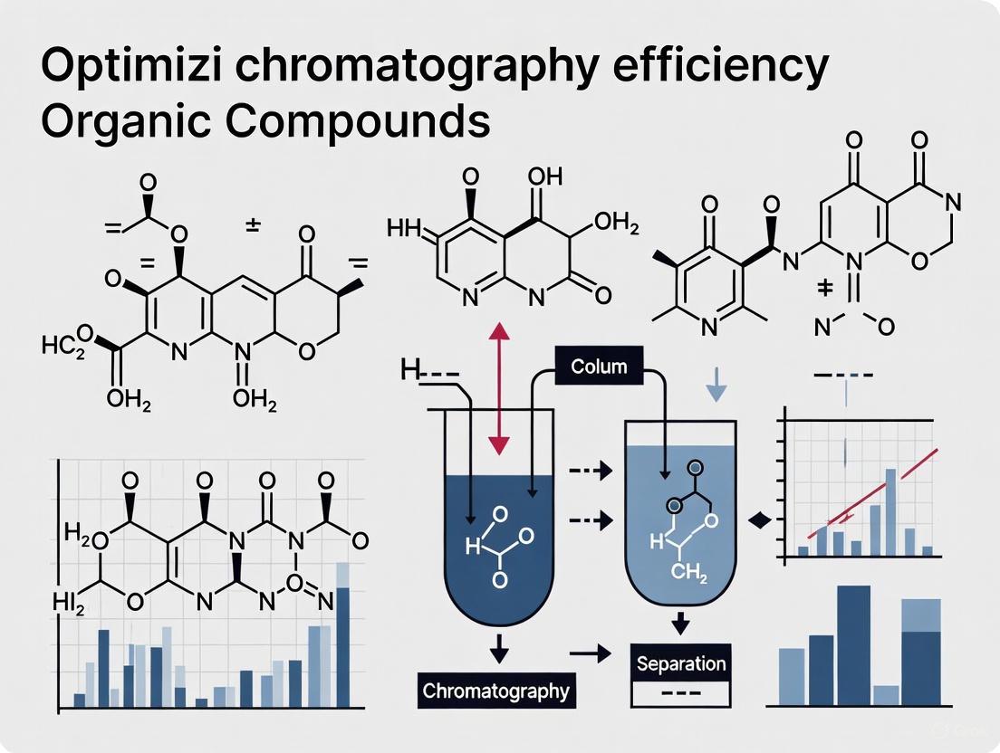
In the pursuit of optimizing chromatography for organic compounds research, scientists and drug development professionals must master the fundamental principles governing separation performance. Chromatographic efficiency quantifies how well a column can separate mixture components into distinct, sharp peaks, directly impacting the resolution, sensitivity, and speed of analytical methods. Three core concepts form the foundation for understanding and optimizing this efficiency: plate count (N), which measures column efficiency; resolution (Rs), which quantifies the degree of separation between two peaks; and the van Deemter equation, which describes how experimental conditions affect efficiency [1] [2]. A thorough grasp of these interrelated concepts enables researchers to systematically develop robust, high-performance chromatographic methods for complex organic mixtures, ultimately leading to more reliable identification and quantification in pharmaceutical and natural product analysis.
Theoretical Plates and Plate Count
The concept of theoretical plates (N) is a cornerstone for measuring column efficiency in chromatography. Adapted from fractional distillation theory by A.J.P. Martin and R.L.M. Synge—work that earned the Nobel Prize in Chemistry in 1952—the theoretical plate model provides a quantitative measure of column performance [2]. In practical terms, the number of theoretical plates is directly related to peak sharpness; a high plate number signifies an efficient column capable of producing narrow, well-resolved peaks, whereas a low plate number results in broad, overlapping peaks and poor separation [2].
The plate height, or Height Equivalent to a Theoretical Plate (HETP), normalizes efficiency relative to column length (L), allowing for meaningful comparisons between different systems. It is defined as ( H = L / N ) [3]. A smaller H-value indicates greater efficiency per unit column length. Plate count can be determined experimentally from a chromatogram using one of several related equations. The most common, approved by the International Union of Pure and Applied Chemistry (IUPAC), uses the retention time (( tR )) and the peak width at its base (( W{base} )) or at half height (( W_{1/2} )) [3]. Table 1 summarizes the key formulas for calculating column efficiency.
Table 1: Formulas for Calculating Column Efficiency and Resolution
| Parameter | Formula | Description |
|---|---|---|
| Plate Count (N) | ( N = 16 \times \left( \frac{tR}{W{base}} \right)^2 ) | Measures column separation efficiency [3] [4]. |
| ( N = 8 \times \ln(2) \times \left( \frac{tR}{W{1/2}} \right)^2 ) | Alternative calculation using peak width at half height [3]. | |
| Plate Height (H) | ( H = \frac{L}{N} ) | Height Equivalent to a Theoretical Plate; lower is better [3] [5]. |
| Resolution (Rₛ) | ( Rs = 1.18 \times \frac{t{R2} - t{R1}}{W{1, h/2} + W_{2, h/2}} ) | Measures the degree of separation between two adjacent peaks [6]. |
Beyond its role in method development, plate count serves as a vital diagnostic tool for monitoring column health over time. A declining plate count often provides the earliest warning of column degradation—such as void formation, contamination, or stationary phase damage—alerting the scientist to potential issues before resolution is critically compromised [2].
Figure 1: Fundamental Causes of Chromatographic Peak Broadening. The diagram illustrates the three primary band-broadening phenomena described by the van Deemter equation and the factors that influence them [1] [7].
The van Deemter Equation
The van Deemter equation, developed in 1956, provides a comprehensive theoretical framework for understanding the band-broadening phenomena that limit column efficiency [1] [3]. It mathematically expresses the plate height (H) as a function of the linear velocity of the mobile phase (u), allowing scientists to identify the optimal flow rate for maximum efficiency. The equation is a hyperbolic function that accounts for the physical, kinetic, and thermodynamic properties of a separation [3].
The classic form of the van Deemter equation is: HETP = A + B/u + C·u Where:
- HETP is the Height Equivalent to a Theoretical Plate, a measure of the resolving power of the column [m] [3] [5].
- u is the linear velocity of the mobile phase [m s⁻¹] [3].
- A is the eddy diffusion term, related to channeling through a non-ideal packing [m] [1] [3].
- B is the longitudinal diffusion coefficient of the eluting particles [m² s⁻¹] [3].
- C is the resistance to mass transfer coefficient between mobile and stationary phases [s] [3].
In-Depth Analysis of the A, B, and C Terms
The A-Term (Eddy Diffusion): This term accounts for the multiple, variable pathways that analyte molecules can take through a packed column due to the non-uniform size and arrangement of packing particles [1]. Molecules following shorter paths elute faster, while those trapped in longer, more tortuous paths elute later, resulting in peak broadening. The A term can be minimized by using uniformly packed, small, spherical particles which create a more consistent flow path [1]. In open tubular capillary columns, the A term is zero because there is no packing [3].
The B-Term (Longitudinal Diffusion): This term describes the natural tendency of analyte molecules to diffuse from regions of high concentration to low concentration along the longitudinal axis of the column [1] [7]. This effect is most pronounced at low mobile phase velocities because molecules spend more time in the column, allowing diffusion more time to spread the peak. To mitigate longitudinal diffusion, higher flow rates can be employed, reducing the time analytes spend in the column [1]. The B term is directly related to the diffusion coefficient of the solute in the mobile phase (( D_M )) [7].
The C-Term (Resistance to Mass Transfer): This term represents the finite time required for analyte molecules to equilibrate (partition) between the stationary and mobile phases [1] [5]. If the mobile phase moves too quickly, some molecules in the mobile phase are swept forward before they can enter the stationary phase, while molecules in the stationary phase lag behind. This disequilibrium causes peak broadening. Factors influencing this term include the thickness of the stationary phase and the diffusion coefficients of the analytes in both phases [1] [7]. The C term becomes the dominant source of band broadening at high flow rates.
The van Deemter Curve and Optimal Velocity
When the plate height (H) is plotted against the linear velocity (u), it typically produces a U-shaped curve, known as a van Deemter plot [5]. The lowest point on this curve represents the optimal linear velocity (( u{opt} ))), where the combined band-broadening effects are minimized, and column efficiency is maximized [1] [5]. The optimum velocity can be derived mathematically from the van Deemter equation and is given by ( u{opt} = \sqrt{B/C} ) [3].
The shape of the van Deemter curve is system-dependent. For instance, Supercritical Fluid Chromatography (SFC) often exhibits flatter C-term regions because supercritical carbon dioxide has low viscosity and a high diffusion coefficient. This allows SFC to be operated at higher linear velocities than HPLC without significant loss of efficiency, leading to faster analysis times [8] [5].
Resolution: The Ultimate Goal of Separation
While plate count measures column efficiency, resolution (Rₛ) is the practical metric that quantifies the success of a separation between two specific analytes. It defines how completely two adjacent peaks are separated from one another [6]. A higher resolution value indicates a better separation. The formula for resolution, as provided by Wyatt's ASTRA software, is ( Rs = 1.18 \times (t{R2} - t{R1}) / (W{1, h/2} + W{2, h/2}) ), where ( tR ) is retention time and ( W_{h/2} ) is the peak width at half height [6].
Resolution is the critical parameter that determines whether a method is fit for purpose, as it directly impacts the ability to accurately identify and quantify individual components in a mixture. Figure 2 illustrates the calculation of resolution and its dependence on efficiency, selectivity, and retention.
Figure 2: Workflow for Calculating and Interpreting Chromatographic Resolution. The diagram outlines the process of determining resolution (Rₛ) from a chromatogram and how to interpret the resulting value to judge the quality of a separation [6].
Experimental Protocols for Efficiency Optimization
Protocol 1: Determining the van Deemter Curve for a Reversed-Phase HPLC System
This protocol provides a step-by-step methodology for empirically determining the optimal flow rate for a given chromatographic system using the principles of the van Deemter equation.
- Column Characterization: Record the column dimensions (length and internal diameter) and stationary phase information (particle size, chemistry).
- Mobile Phase Preparation: Prepare a degassed, isocratic mobile phase suitable for the column and a test analyte. A common example is 50:50 methanol/water for a reversed-phase C18 column.
- Test Solution: Prepare a standard solution of a stable, well-retained, and pure analyte (e.g., caffeine for reversed-phase) at a low concentration to avoid overloading the column.
- Instrumental Parameters:
- Detector: UV-Vis, wavelength set to the λₘₐₓ of the analyte.
- Column Temperature: Set and maintain a constant temperature (e.g., 25°C).
- Injection Volume: Keep small and consistent (e.g., 5-10 µL).
- Data Acquisition:
- Set the flow rate to an initial low value (e.g., 0.2 mL/min).
- Inject the standard and record the chromatogram.
- Accurately record the retention time (( tR )) and the peak width at half height (( W{1/2} )) of the analyte peak.
- Calculate the linear velocity (u) from the flow rate (F) and the column's cross-sectional area (A): ( u = F / A ).
- Incrementally increase the flow rate (e.g., 0.4, 0.6, 0.8, 1.0, 1.2 mL/min) and repeat the injection and measurement at each step.
- Data Analysis:
- For each flow rate/velocity, calculate the plate count (N) using ( N = 8 \times \ln(2) \times (tR / W{1/2})^2 ) [3].
- Calculate the plate height (H) for each point using ( H = L / N ).
- Plot H (y-axis) against u (x-axis) to generate the van Deemter curve.
- Identify the optimum linear velocity (( u_{opt} )) at the minimum of the curve.
Protocol 2: Monitoring Column Performance and Health Over Time
This protocol is used for routine column diagnostics and to track performance degradation, which is critical for maintaining data integrity in long-term studies.
- Establish a Baseline: When a new column is installed, run a standardized test mixture under predefined, isocratic conditions to establish its initial performance baseline. Record the plate count (N), peak asymmetry factor (As), and backpressure.
- Standard Test Mixture: Use a mixture of compounds that are well-characterized and appropriate for the column chemistry. For a C18 column, this might include uracil (for dead time, t₀) and a few small, neutral aromatic compounds like toluene or naphthalene.
- Routine Monitoring: At regular intervals (e.g., weekly or every 100 injections), under the same standardized conditions, inject the test mixture.
- Data Collection and Calculation:
- For each key peak in the test mixture, calculate the plate count (N) and asymmetry factor (As) using the chromatography data system (CDS) software. As defined by the US Pharmacopeia, the asymmetry factor is calculated at 10% of the peak height [6].
- Record the system backpressure.
- Performance Tracking:
The Scientist's Toolkit: Essential Reagents and Materials
Table 2: Key Research Reagent Solutions for Chromatographic Analysis
| Item | Function / Purpose |
|---|---|
| Chromatography Column | The heart of the separation; its dimensions (length, internal diameter), particle size, and stationary phase chemistry (e.g., C18, silica, HILIC) define the system's intrinsic efficiency and selectivity [1] [2]. |
| Mobile Phase Solvents & Modifiers | High-purity solvents (e.g., water, acetonitrile, methanol) are used to create the eluent. Modifiers (e.g., acids, bases, salts) adjust pH and ionic strength to control retention, selectivity, and peak shape [1]. |
| Supercritical CO₂ | Serves as the primary, non-toxic, and reusable mobile phase in Supercritical Fluid Chromatography (SFC), offering high diffusion coefficients and low viscosity for faster, greener separations [8] [5]. |
| Micellar Eluents | Used in Micellar Liquid Chromatography (MLC) as a green alternative to reduce consumption of toxic organic solvents while maintaining efficient separations [8]. |
| Natural Deep Eutectic Solvents (NADES) | Emerging as green, biodegradable, and low-toxicity alternatives for sample preparation and extraction, aligning with green chemistry principles [8]. |
| Standard Test Mixtures | Solutions of known, pure analytes (e.g., caffeine, naphthalene, alkyl phenones) used for system suitability testing, column performance validation, and generating van Deemter curves [6]. |
Advanced Applications and Considerations
Preparative vs. Analytical Chromatography
The role of efficiency and plate count differs significantly between analytical and preparative chromatography. In analytical chromatography, the goal is detection and quantification, so high plate counts are paramount for achieving maximum resolution [4]. In preparative chromatography, where the goal is to isolate large quantities of material, loading capacity becomes the primary concern. Interestingly, higher efficiency columns (typically packed with smaller particles) overload more quickly than columns packed with larger particles. As shown in Equation 2 from Biotage, loading capacity (Mi) is inversely proportional to the square root of the plate count (n): ( M_i \propto 1 / n^{1/2} ) [4]. This is one reason why larger particle sizes (e.g., 20-40 µm) are often preferred in flash chromatography, as they offer a better balance between separation performance and loading capacity [4].
Modern Variations of the van Deemter Equation
The original van Deemter equation has been expanded and modified to account for different column geometries and modern stationary phases. The Golay equation applies to open tubular capillary columns, where the A term is zero due to the lack of packing [3]. The Rodrigues equation extends the model to describe the efficiency of beds packed with permeable (large-pore) particles, incorporating a function of the intraparticle Peclet number [3]. Furthermore, contemporary research accounts for high-pressure conditions that create longitudinal temperature and pressure gradients, altering diffusion coefficients along the column and requiring more complex forms of the C-term in the van Deemter equation [1].
A deep and practical understanding of plate count, resolution, and the van Deemter equation is indispensable for any researcher aiming to optimize chromatographic efficiency in the analysis of organic compounds. These concepts are not isolated theories but are powerfully interconnected tools. The plate count provides a measure of intrinsic column performance, the van Deemter equation offers a roadmap for optimizing operational parameters like flow rate, and resolution is the ultimate, practical measure of a successful separation. By systematically applying the principles and experimental protocols outlined in this application note—from generating van Deemter curves to routine column health monitoring—scientists and drug development professionals can develop more robust, efficient, and reliable chromatographic methods. This systematic approach ultimately accelerates research and ensures the highest quality data in pharmaceutical and natural product analysis.
The global chromatography market is experiencing significant growth, propelled by robust demand from key sectors including biopharmaceuticals, generics, and environmental testing. For researchers and drug development professionals, optimizing chromatography efficiency is paramount to navigating this expanding landscape. This involves leveraging advanced instrumentation, innovative column technologies, and refined methodologies to meet stringent regulatory requirements and the need for high-throughput analysis. This article provides a detailed analysis of the current market drivers and presents structured application notes and experimental protocols to enhance chromatographic workflows for organic compounds research, with a focus on practical implementation.
The chromatography market is demonstrating strong growth, with specific segments and regions showing particularly high activity. The data below summarizes the current market size and projections.
Table 1: Global Chromatography Market Size and Projections
| Market Segment | Base Year (2024) Market Size | 2025 Market Size | Projected 2030 Market Size | CAGR (2025-2030) |
|---|---|---|---|---|
| Pharmaceuticals & Biotechnology | $12.3 billion [9] | $13.3 billion [9] | $19.8 billion [9] | 8.4% [9] |
This growth is unevenly distributed across different regions and technology types. The following table breaks down the dominant segments.
Table 2: Market Dominance by Region and Technology
| Category | Dominant Region/Technology | Market Share / Key Statistic |
|---|---|---|
| Regional Share | North America [9] | Accounts for 45% of the global market [9] |
| Technology Segment | Liquid Chromatography [9] | Dominant technology through 2030 [9] |
Application Note 1: Biopharmaceutical Characterization
Background and Objective
The biopharmaceutical market's surge, driven by innovations like monoclonal antibodies (mAbs), cell and gene therapies, and peptide-based drugs such as GLP-1 agonists, demands highly efficient purification and characterization techniques [10] [9]. Chromatography is indispensable for ensuring the purity, stability, and efficacy of these complex molecules by characterizing Critical Quality Attributes (CQAs) [9] [11]. This application note details a protocol for the rapid analysis of a monoclonal antibody using advanced liquid chromatography, reducing analysis time from hours to minutes while maintaining resolution [11].
Experimental Protocol
Method: Rapid High-Performance Liquid Chromatography (HPLC) for mAb Charge Variant Analysis [11]
Workflow Overview:
Step-by-Step Procedure:
- Sample Preparation: Dilute the monoclonal antibody sample to a concentration of 1 mg/mL using a compatible buffer such as 20 mM phosphate buffer, pH 7.0. Centrifuge at 10,000 x g for 5 minutes to remove any particulates [11].
- Column Selection: Utilize a reversed-phase C18 column with superficially porous particles (e.g., 2.7 μm particle size) housed in inert (metal-free) hardware. This configuration enhances peak shape, improves analyte recovery for metal-sensitive species, and provides the efficiency needed for rapid separations [11] [12].
- Instrument Parameters:
- System: Ultra-High-Performance Liquid Chromatography (UHPLC) system capable of operating at pressures up to 1000 bar.
- Mobile Phase: Employ a gradient method.
- Mobile Phase A: 0.1% Trifluoroacetic acid (TFA) in water.
- Mobile Phase B: 0.1% TFA in acetonitrile.
- Gradient: Ramp from 20% B to 60% B over 5 minutes.
- Flow Rate: 0.5 mL/min.
- Column Temperature: 50°C.
- Detection: UV absorbance at 280 nm [11].
- Data Analysis: Use integrated software to identify and quantify the various charge variants (acidic, main, and basic species) based on peak area and retention time. The method should be validated for specificity, linearity, and precision [11].
Application Note 2: Purification for Generics and APIs
Background and Objective
The growing market for generics and biosimilars requires robust, cost-effective methods to confirm molecular similarity, purity, and bioequivalence [9]. A key challenge is the purification of highly polar Active Pharmaceutical Ingredients (APIs), which often exhibit poor retention and peak shape on traditional silica-based columns [13]. This protocol describes an Ion-Assisted Chromatography technique that improves the purification of polar amines and peptide APIs without the need for expensive specialized purification materials [13].
Experimental Protocol
Method: Normal-Phase Chromatography with Ionic Additives for Polar API Purification [13]
Workflow Overview:
Step-by-Step Procedure:
- Sample Preparation: Prepare the crude reaction mixture containing the polar organic compound (e.g., an amine salt or peptide) by dissolving it in a minimal volume of an appropriate solvent like dichloromethane or methanol [13].
- Column Packing: Pack a standard glass chromatography column with silica gel as the stationary phase. This method is effective with common, inexpensive silica, unlike other techniques that require costly alternatives [13].
- Mobile Phase Modification: This is the critical step. Add calcium chloride (CaCl₂) to the aqueous component of your normal-phase mobile phase system at a concentration of 10-50 mM. The calcium ions interact with the polar functional groups, improving separation efficiency and peak shape [13].
- Elution and Fraction Collection: Run the chromatography using a standard normal-phase elution protocol (e.g., a gradient of methanol in dichloromethane). Collect fractions based on UV detection or TLC monitoring [13].
- Analysis of Fractions: Analyze the collected fractions using a standard analytical HPLC method to assess the purity of the isolated API. Compare the results with those from a purification run without CaCl₂ to demonstrate the improvement in resolution and yield [13].
The Scientist's Toolkit: Key Research Reagent Solutions
Optimizing chromatography requires careful selection of materials. The following table details key solutions for modern challenges.
Table 3: Essential Reagents and Materials for Chromatography Optimization
| Item Name | Function/Application | Key Benefit |
|---|---|---|
| Superficially Porous Particle (SPP) Columns [12] | High-resolution separation of small molecules and peptides in RPLC. | Provides high efficiency and throughput with lower backpressure compared to fully porous particles. |
| Inert/Bioinert Hardware Columns [12] | Analysis of metal-sensitive compounds (e.g., phosphorylated molecules, peptides, chelating PFAS). | Prevents analyte adsorption to metal surfaces, improving peak shape and recovery. |
| Ion-Pairing Reagent Free Columns for Oligonucleotides [12] | Separation of oligonucleotides without the use of ion-pairing reagents. | Simplifies method development and enhances compatibility with mass spectrometry. |
| Calcium Chloride (CaCl₂) Additive [13] | Purification of highly polar organic compounds (amines, peptides) in normal-phase chromatography. | Enables use of inexpensive silica gel for challenging separations, reducing costs. |
| Green Solvents (e.g., CO₂ for SFC, NADES) [8] | Sustainable extraction and analysis of natural products. | Reduces consumption of toxic organic solvents, aligning with green chemistry principles. |
Application Note 3: Environmental Testing of Contaminants
Background and Objective
Environmental monitoring, particularly for persistent organic pollutants like per- and polyfluoroalkyl substances (PFAS), is a major driver of chromatography demand [10]. Regulatory agencies mandate strict testing protocols, such as EPA Method 8270E, which defines the determination of semivolatile organic compounds in solid and water matrices using gas chromatography/mass spectrometry (GC-MS) [14]. This protocol outlines a compliant workflow for the analysis of semivolatile organics in environmental samples.
Experimental Protocol
Method: Analysis of Semivolatile Organic Compounds by GC-MS per EPA Method 8270E [14]
Workflow Overview:
Step-by-Step Procedure:
- Sample Collection and Extraction:
- Extract Cleanup and Concentration: Cleanup the raw extract to remove interfering co-extractives using techniques such as gel permeation chromatography (GPC) or silica gel cleanup. Concentrate the purified extract to a final volume of 1.0 mL using a gentle stream of nitrogen [14].
- GC-MS Analysis:
- Instrument: GC system coupled with a mass spectrometric detector.
- Column: Fused-silica capillary column (e.g., 30 m length x 0.25 mm ID, coated with 0.25 μm of 5% diphenyl/95% dimethyl polysiloxane).
- Oven Program: Temperature program from 40°C (hold 2 min) to 320°C at a rate of 10°C/min.
- Injector: Use a splitless injection mode at 250°C.
- MS Detection: Operate in electron impact (EI) ionization mode at 70 eV. Use selected ion monitoring (SIM) for quantitative analysis [14].
- Calibration and Quantification: Establish a multi-point calibration curve using certified analytical standards for the target semivolatile compounds. Quantify the analytes in the sample extracts by comparing their response to the calibration curve. Include quality control samples (blanks, duplicates, and matrix spikes) to ensure data integrity [14].
The chromatography sector is robust, fueled by critical applications in biopharmaceuticals, generics, and environmental testing. Success in this field hinges on the adoption of optimized methods and technologies. The application notes and protocols provided here—encompassing rapid HPLC for biologics, innovative ion-assisted purification for APIs, and rigorous GC-MS for environmental compliance—offer researchers and scientists a practical framework to enhance analytical efficiency, ensure data quality, and maintain regulatory alignment in their organic compounds research.
Liquid Chromatography-Mass Spectrometry (LC-MS) and Gas Chromatography-Mass Spectrometry (GC-MS) represent cornerstone hyphenated techniques in modern analytical laboratories. This application note provides a detailed comparison of these platforms, framed within the context of optimizing chromatography efficiency for organic compound research. We summarize core technical differences, present current utilization trends, introduce novel sample preparation products, and provide detailed protocols for method optimization to support researchers and drug development professionals.
Core Technique Comparison and Current Trends
GC-MS and LC-MS are complementary techniques whose selection depends primarily on the physicochemical properties of the target analytes. The following table summarizes their fundamental characteristics.
Table 1: Core Differences Between GC-MS and LC-MS
| Feature | GC-MS | LC-MS |
|---|---|---|
| Best For | Volatile, thermally stable, and non-polar compounds [16] | Polar, large, or thermally unstable molecules (e.g., peptides, proteins, metabolites) [16] |
| Separation Mechanism | Gas chromatography using an inert gas mobile phase [16] | Liquid chromatography using liquid solvents as the mobile phase [16] |
| Sample Preparation | Often requires derivatization for non-volatile analytes [16] | Usually minimal preparation; no derivatization typically needed [16] |
| Ionization Source | Electron Ionization (EI) or Chemical Ionization (CI) [17] | Electrospray Ionization (ESI) or Atmospheric Pressure Chemical Ionization (APCI) [17] [18] |
| Typical Applications | Residual solvent analysis, impurity profiling, pesticide monitoring [19] [16] | Biomarker quantification, peptide sequencing, metabolomics, therapeutic drug monitoring [17] [16] |
Analysis of the scientific literature reveals distinct publication trends. From 1995–2023, the yearly publication rate for GC-MS was nearly linear at an estimated 3,042 articles per year, while for LC-MS (1997–2023) it was higher, at 3,908 articles per year (LC-MS/GC-MS ratio of 1.3:1). This trend continued into 2024, with an estimated ratio of 1.5:1, indicating the growing utilization of LC-MS in life sciences research [17]. These techniques are used worldwide, with leading countries including China, the United States, Germany, and Japan [17].
Advanced Application Notes: Sample Preparation for 2024-2025
Innovations in sample preparation are critical for enhancing chromatographic efficiency and data quality. The following novel products address key challenges in analyzing complex matrices.
Table 2: New Sample Preparation Products (2024-2025)
| Product Name | Vendor | Application | Key Feature and Benefit |
|---|---|---|---|
| Captiva EMR PFAS Food Cartridge [19] | Agilent Technologies | PFAS analysis in food matrices (EPA, FDA methods) | Pass-through cleanup that eliminates manual QuEChERS steps, saving time and reducing waste. |
| Resprep PFAS SPE [19] | Restek | PFAS in aqueous/solid samples (EPA Method 1633) | Dual-bed SPE with filter aid to minimize clogging and avoid wool packing. |
| InertSep WAX FF/GCB [19] | GL Sciences | PFAS analysis using EPA Method 1633 | High-purity sorbents and optimized particle size minimize contamination and improve permeability. |
| Captiva EMR Mycotoxins [19] | Agilent Technologies | Multiclass mycotoxin analysis in food/feed | Simplifies workflow by eliminating multiple extraction protocols for different mycotoxin classes. |
| Samplify Automated System [19] | Sielc Technologies | Unattended, periodic liquid sampling | Enables adjustable sampling volumes, automatic mixing, dilution, and probe cleaning for high reproducibility. |
Detailed Experimental Protocols
Protocol 3.1: Solid-Phase Extraction (SPE) for PFAS in Aqueous Samples using EPA Method 1633
This protocol utilizes the Restek Resprep PFAS SPE cartridge [19].
I. Research Reagent Solutions Table 3: Essential Materials for PFAS SPE
| Item | Function |
|---|---|
| Resprep PFAS SPE Cartridge | Dual-bed cartridge for specific extraction and cleanup of PFAS from aqueous matrices. |
| Methanol (HPLC-grade) | For cartridge conditioning and analyte elution. |
| Ammonium Acetate Buffer | Provides the appropriate ionic strength and pH for optimal analyte retention. |
| Water (HPLC-grade) | For sample dilution and cartridge washing. |
| Vacuum Manifold | Provides positive pressure for consistent and efficient flow through the SPE cartridge. |
II. Step-by-Step Workflow
- Conditioning: Pass 5 mL of methanol through the SPE cartridge, followed by 5 mL of reagent water. Do not allow the sorbent bed to dry out.
- Sample Loading: Load the acidified aqueous sample (typically 250 mL) onto the cartridge at a steady flow rate of 5-10 mL per minute.
- Washing: After sample loading, wash the cartridge with 5 mL of a 25 mM ammonium acetate buffer (pH 4) to remove interfering compounds.
- Drying & Elution: Apply a vacuum for 10 minutes to dry the sorbent completely. Elute the PFAS analytes into a collection tube using 5 mL of methanol.
- Concentration & Reconstitution: Evaporate the eluent to dryness under a gentle stream of nitrogen. Reconstitute the dried extract in 0.5 mL of methanol or a compatible LC-MS initial mobile phase for analysis.
Protocol 3.2: Optimizing LC-MS/MS Sensitivity using Design of Experiments (DOE)
This protocol uses a Definitive Screening Design (DSD) to efficiently optimize multiple MS parameters simultaneously, a statistically superior approach to one-factor-at-a-time (OFAT) testing [20].
I. Research Reagent Solutions Table 4: Key Materials for LC-MS/MS Optimization
| Item | Function |
|---|---|
| Standard Solutions | Pure analyte and internal standard for consistent signal measurement. |
| LC-MS Mobile Phases | e.g., Water and methanol/acetonitrile, both with volatile additives (e.g., formic acid, ammonium acetate). |
| Statistical Software | Software capable of DOE (e.g., JMP, Minitab, or R with appropriate packages). |
II. Step-by-Step Workflow
- Define Factors and Ranges: Select key MS parameters and their high/low values based on instrument capabilities and preliminary data.
- Factors: Capillary Voltage (e.g., 2.5 - 4.0 kV), Drying Gas Temperature (e.g., 250 - 350 °C), Nebulizer Gas Flow (e.g., 30 - 50 psi), Collision Energy (a range appropriate for the analyte).
- Create Experimental Design: Use a DSD, which requires only 2k+1 experiments (where k is the number of factors) to screen many factors and identify critical interactions [20].
- Randomize and Execute: Run the experiments in a randomized order to protect against unknown biases and systematic error [20].
- Measure Response and Model Data: The primary response variable is the Signal-to-Noise Ratio of the analyte. Fit the data to a statistical model to identify significant factors and their optimal settings.
- Verify the Optimum: Perform a confirmation run using the predicted optimal parameters to validate the model's accuracy.
The workflow for this systematic optimization is outlined below.
Critical Method Optimization Parameters
Beyond systematic DOE, several individual parameters require careful attention during method development.
- Ionization Mode Selection: While electrospray ionization (ESI) is best for polar/ionizable compounds and atmospheric pressure chemical ionization (APCI) for less polar molecules, screen all available techniques and both polarity modes for unexpected optimal response [18].
- Capillary Voltage Optimization: This is a frequently overlooked parameter that majorly impacts ionization efficiency and reproducibility. Avoid non-ideal spray modes that cause signal variability [18].
- Mobile Phase and Additives: Adjust the eluent pH to be at least 1 unit above (for acids) or below (for bases) the analyte pKa to ensure ionization, boosting sensitivity orders of magnitude. Use volatile buffers (e.g., ammonium formate) and avoid non-volatile ion-pairing reagents like trifluoroacetic acid (TFA) [18].
- Collision Energy and Dwell Time: In MS/MS analysis, optimize collision energy for each transition. Ensure sufficient dwell time between measuring ion transitions to avoid "cross-talk" that compromises quantitative accuracy [18].
The logical relationship between sample properties and the optimal choice of chromatographic technique is summarized in the following decision pathway.
Current Market Landscape and Instrumentation Trends for 2025
The global chromatography instrumentation market is demonstrating robust growth, driven by escalating demand from the pharmaceutical, biotechnology, and environmental testing sectors. This expansion is quantified in the following data summaries.
Table 1: Global Chromatography Instrumentation Market Size and Growth (2024-2032)
| Metric | Value / Forecast | Time Period | Data Source |
|---|---|---|---|
| Market Size (2024) | USD 12.3 Billion | 2024 | [21] |
| Market Size (2025E) | USD 10.31 Billion | 2025 | [22] |
| Market Size (2031F) | USD 18.8 Billion | 2031 | [21] |
| Market Size (2032F) | USD 14.82 Billion | 2032 | [22] |
| CAGR (2024-2031) | 6.1% | 2024-2031 | [21] |
| CAGR (2025-2032) | 5.32% | 2025-2032 | [22] |
Table 2: Chromatography Instrumentation Market Share by Technology (2025)
| Technology | Market Share (%) | Key Application Drivers |
|---|---|---|
| Liquid Chromatography (LC) | 50.2% | Pharmaceuticals, Biotechnology, Clinical Research [22] [21] |
| Gas Chromatography (GC) | 33.0% | Environmental Monitoring, Petrochemicals, Food & Beverage [23] |
| Supercritical Fluid Chromatography (SFC) | 9.0% | Chiral Separations, Green Chemistry [23] |
| Thin-Layer Chromatography (TLC) | 7.0% | Academic Research, Low-Cost QC [23] |
Key market drivers include rising pharmaceutical research and development, which accounts for over 52% of global chromatography application demand [23]. Stringent regulatory requirements for drug approval and environmental monitoring, such as the US EPA's Method 1633 for PFAS analysis, further propel market growth [24]. The market faces restraints, including the high upfront and maintenance costs of advanced systems, which can exceed USD 500,000, limiting adoption in smaller laboratories [24] [23].
Key Market Trends and Strategic Directions
Dominance of Liquid Chromatography and Technological Evolution
Liquid chromatography continues to lead the market, with Ultra-High-Performance Liquid Chromatography (UHPLC) becoming the standard for high-throughput and high-sensitivity applications. Its dominance is anchored in its unmatched versatility for analyzing complex mixtures, from small molecules to large biologics, in drug development and quality control [22] [21]. Recent trends focus on platforms that offer higher pressure limits (exceeding 1,300 bar), embedded self-diagnostic sensors, and AI-driven gradient optimization to enhance peak capacity and reduce solvent consumption by up to 65% [24].
Green Chromatography and Sustainability
A significant shift toward environmentally sustainable practices is underway. Key developments include:
- Supercritical Fluid Chromatography (SFC): Gaining traction as a green alternative that utilizes supercritical CO₂ as the primary mobile phase, drastically reducing the consumption of toxic organic solvents [8] [23].
- Solvent Reduction Strategies: Widespread adoption of miniaturized systems, microextraction techniques, and low-flow UHPLC to minimize waste generation [8] [23].
- Alternative Mobile Phases: Exploration and application of Micellar Liquid Chromatography (MLC) and Natural Deep Eutectic Solvents (NADES) for sample preparation and analysis, offering biodegradability and low toxicity [8].
Automation, AI, and Digital Integration
Laboratories are increasingly adopting smart, connected instruments to improve efficiency and data integrity.
- AI-Powered Workflows: Approximately 30-34% of top-tier labs are implementing AI-enabled software for real-time method optimization, automated peak integration, and predictive maintenance, reducing analytical errors by nearly 20% [24] [23].
- Cloud-Based Platforms: Around 25% of large-scale labs use cloud-connected chromatography systems for remote monitoring, data management, and global collaboration, facilitating regulatory compliance and data traceability [23].
Portable and Miniaturized Systems
The demand for on-site analysis is driving the growth of portable and miniaturized chromatography instruments. Over 15,000 portable units were deployed globally in 2023, particularly for environmental testing and forensic applications, offering rapid results outside the traditional laboratory setting [23].
Regional Market Analysis
Table 3: Regional Market Share and Growth Trends
| Region | Market Share (2025) | Key Growth Drivers |
|---|---|---|
| North America | 38.3% - 38.7% | Well-established pharmaceutical & biotech sector, stringent FDA regulations, high R&D investment [22] [23] |
| Europe | ~29% | Strict environmental (REACH) and solvent usage regulations, strong pharmaceutical manufacturing base [24] [23] |
| Asia-Pacific | 25.2% (Fastest Growing) | Rapid industrialization, expanding pharmaceutical manufacturing (especially in China & India), government healthcare initiatives [22] [21] |
Experimental Protocols for Modern Chromatography
Protocol: Rapid GC-MS Profiling of Small Organic Molecules in Biomass Pyrolysis
This industry-ready method is ideal for process optimization and can be adapted for various volatile organic compound analyses [25].
4.1.1 Research Reagent Solutions
| Item | Function / Specification |
|---|---|
| DB-WAX-UI Column | A polar stationary phase for separating volatile acids and alcohols. |
| High-Purity Helium (He) or Hydrogen (H₂) | Mobile phase carrier gas. Hydrogen is increasingly used due to helium supply shortages. |
| Standard Solutions | Analytical standards of formaldehyde, methanol, acetic acid, and formic acid for calibration. |
| Derivatization Reagent | (If needed) To increase volatility of target analytes. |
| Appropriate Solvent | e.g., Methanol or Hexane, for preparing standard and sample solutions. |
4.1.2 Workflow Diagram
4.1.3 Detailed Methodology
- Sample Preparation: Perform simple sample preparation, which may involve filtration or dilution of the collected biomass pyrolysis condensate. The method emphasizes minimal and straightforward preparation [25].
- Instrumental Setup:
- GC: Configured with a DB-WAX-UI column (30 m length, 0.25 mm internal diameter, 0.25 µm film thickness recommended).
- Oven Program: Optimize a temperature ramp to achieve baseline separation of all target analytes (formaldehyde, methanol, acetic acid, formic acid) in under 15 minutes.
- MS: Operate with an electron impact (EI) ion source.
- Data Acquisition:
- Selected Ion Monitoring (SIM): Acquire data for target quantitation of specific analytes to achieve high sensitivity. Reported Instrumental LODs: Formaldehyde (0.09 µg mL⁻¹), Methanol (0.04 µg mL⁻¹), Acetic Acid (0.02 µg mL⁻¹), Formic Acid (0.03 µg mL⁻¹) [25].
- Full Scan Mode (m/z 40-300): Acquire data concurrently for untargeted profiling and detection of unknown compounds in the sample.
- Validation: The method demonstrates high repeatability, with intra-day and inter-day precision (RSD) better than 5% and 12%, respectively [25].
Protocol: UHPLC-MS Method Development for Complex Organic Mixtures
This protocol outlines a modern approach for separating and analyzing complex samples, such as natural products or pharmaceutical impurities.
4.2.1 Research Reagent Solutions
| Item | Function / Specification |
|---|---|
| UHPLC System | Capable of operating at pressures > 1,000 bar. |
| C18 Column with Small Particle Size | e.g., 1.7-1.8 µm particles for high-efficiency separations. |
| Mass Spectrometer Detector | e.g., Q-TOF or Tandem Quadrupole MS for identification and quantification. |
| Mobile Phase A | High-purity water with 0.1% Formic Acid. |
| Mobile Phase B | Methanol or Acetonitrile with 0.1% Formic Acid. |
| Biocompatible Columns/Components | (For biomolecules) With MaxPeak Premier coating or similar to minimize metal adsorption [24]. |
4.2.2 Workflow Diagram
4.2.3 Detailed Methodology
- Sample Preparation:
- Dissolve the sample in a solvent compatible with the initial mobile phase composition (e.g., water or a mixture of water and organic solvent).
- Centrifuge or Filter (0.22 µm membrane) to remove particulates that could clog the UHPLC system [26].
- Column Selection: Choose a column chemistry suitable for the target analytes. For reversed-phase analysis of small organic molecules, a C18 column with sub-2µm particles is standard. For sensitive biomolecules, use biocompatible columns to prevent analyte adsorption and improve recovery [24].
- Method Scouting and AI Optimization:
- Utilize software tools that incorporate AI to automatically scout initial gradient conditions and tune them in real-time for optimal peak capacity and minimal solvent use.
- These systems can reduce method development time and solvent consumption by up to 65% [24].
- UHPLC-MS Analysis:
- Perform the separation using the optimized, steep gradient at high pressure.
- Couple the UHPLC system to a high-resolution mass spectrometer for accurate mass identification and quantification.
- Data Analysis: Use advanced software for peak picking, integration, and compound identification against databases.
Market Influence Factors
Advanced Method Development for Complex Organic Molecules
Chromatographic analysis stands as a cornerstone of modern scientific research, particularly in the development and analysis of organic compounds. Achieving optimal separation efficiency requires the careful balancing of three critical parameters: particle size, column length, and flow rate. Individually, each parameter exerts a profound influence on resolution, analysis time, and backpressure. Collectively, they determine the success of any chromatographic method. This application note provides a systematic framework for optimizing these parameters, grounded in fundamental chromatographic theory and contemporary practice, to enhance method development for researchers and drug development professionals.
The interrelationship between these parameters is best understood through the van Deemter equation, which describes the dependence of column efficiency (Height Equivalent to a Theoretical Plate, HETP) on the linear velocity of the mobile phase. The equation models the contributions of eddy diffusion (A-term), longitudinal molecular diffusion (B-term), and mass transfer resistance (C-term) to band broadening [27]. The goal of optimization is to identify the operational conditions that minimize HETP, thereby maximizing efficiency. Smaller particles not only reduce the path for mass transfer but also enable the use of shorter columns while maintaining resolution, which in turn permits adjustments to flow rates for faster analysis without compromising performance [28] [29]. The following sections provide detailed guidance on optimizing each parameter, complete with practical protocols and data-driven recommendations.
Parameter Optimization: Theoretical and Practical Considerations
Particle Size
Particle size refers to the average diameter of the spherical silica or other material that forms the stationary phase packing within the chromatographic column. Modern High-Performance Liquid Chromatography (HPLC) commonly utilizes fully porous particles of 3–5 µm, while Ultra-High-Performance Liquid Chromatography (UHPLC) employs sub-2-µm particles to achieve superior efficiency [28].
Mechanism of Impact: The particle size directly influences the van Deemter C-term (mass transfer resistance). A smaller particle size reduces the distance an analyte must diffuse to enter and exit the pores of the stationary phase. This facilitates faster mass transfer, allowing the analyte to equilibrate more rapidly between the mobile and stationary phases. The result is reduced band broadening, especially at higher flow rates [28] [29]. Furthermore, smaller particles provide a larger surface area per unit volume, enhancing the potential for interactions that improve resolution [28].
Trade-offs and Considerations: The primary trade-off with reduced particle size is a significant increase in backpressure, as described by the Darcy's Law relationship where pressure is inversely proportional to the square of the particle size. This necessitates instrumentation capable of withstanding high pressures (e.g., UHPLC systems). Additionally, columns packed with smaller particles are more susceptible to clogging and require meticulous sample cleaning and the use of high-purity solvents [28]. The particle size distribution (PSD) is also critical; a narrower distribution (e.g., D90/10 value of ~1.1) contributes to more uniform packing and lower eddy diffusion (A-term), further enhancing efficiency and reducing backpressure compared to columns with a wider PSD [29].
Table 1: Impact of Particle Size on Chromatographic Performance
| Particle Size (µm) | Key Advantages | Key Challenges | Typical Application |
|---|---|---|---|
| ≥ 5 | Moderate backpressure, compatible with standard HPLC, cost-effective | Lower efficiency, longer analysis times | Standard QC, simple mixtures |
| 3 - 5 | Balanced efficiency and pressure, widely versatile | Higher backpressure than larger particles | Most routine HPLC analyses |
| < 2 | Highest efficiency, sharper peaks, faster analyses | Very high backpressure, requires UHPLC, clogging risk | Complex mixtures, high-throughput labs |
Column Length
The column length determines the physical path over which the separation occurs, directly impacting the number of theoretical plates (N) available for resolving components in a mixture.
Mechanism of Impact: According to the fundamental relationship, the number of theoretical plates (N) is proportional to column length (L) and inversely proportional to the plate height (H); N = L/H. Therefore, for a given particle size (which influences H), a longer column provides more theoretical plates, potentially yielding higher resolution [30]. This is particularly crucial for separating complex mixtures of small molecules with subtle structural differences.
The "Effective Column Length" for Biomolecules: A critical modern consideration is that the above principle does not hold true for large biomolecules. Due to their "on-off" or "bind-and-elute" elution mechanism in modes like reversed-phase and ion-exchange chromatography, large molecules (e.g., > 5 kDa) are retained only on a very short segment of the column bed. Research by Fekete and Lauber demonstrates that for a 100–150 kDa molecule, the effective column length needed is remarkably short—often just 1–2 cm [31]. Using a column longer than this effective length provides no resolution benefit and only serves to increase analysis time and backpressure. Therefore, for large molecules, short columns (1–3 cm) are recommended, especially when using gradients of less than 20 minutes [31].
Trade-offs and Considerations: For small molecules, increasing column length improves resolution at the cost of longer analysis times and higher backpressure. It also leads to greater solvent consumption, increasing operational costs and environmental impact [30]. The choice of column length must therefore be aligned with the nature of the analytes and the separation goals.
Table 2: Guidelines for Selecting Column Length Based on Application
| Analyte Type | Recommended Column Length | Rationale | Example Separation Modes |
|---|---|---|---|
| Small Molecules (< 1000 Da) | 50 - 150 mm | Longer path provides more theoretical plates for resolving complex mixtures | RPLC, HILIC, Normal Phase |
| Peptides / Small Proteins | 30 - 100 mm | Balance between needed resolution and analysis speed | RPLC, Ion-Exchange |
| Large Biomolecules (> 5 kDa) | 10 - 30 mm | Utilizes the "on-off" elution mechanism; longer columns are ineffective | RPLC, HIC, IEX, HILIC [31] |
Flow Rate
The flow rate of the mobile phase dictates the linear velocity at which analytes travel through the column. It is a key parameter controlling the kinetic aspects of the separation.
Mechanism of Impact: The flow rate's effect on efficiency is precisely described by the van Deemter curve. At very low flow rates, the B-term (longitudinal diffusion) dominates, leading to band broadening. At very high flow rates, the C-term (mass transfer resistance) dominates, as analytes do not have sufficient time to equilibrate between phases. The optimal flow rate is located at the minimum of the van Deemter curve, where the combined contributions of band broadening are minimized [27]. This optimal linear velocity is particle-size dependent; smaller particles have higher optimal linear velocities, allowing for faster separations without a loss of efficiency [32].
Trade-offs and Considerations: Operating above the optimal flow rate sacrifices resolution for speed, a trade-off that may be acceptable in high-throughput screening. Operating below the optimal flow rate maximizes resolution but at the cost of longer analysis times. In practical terms, for flash column chromatography with 25 µm media, a 12-gram column has a practical optimal flow rate around 21 mL/min, while for 50 µm media, it is significantly lower at 6 mL/min [32]. It is also crucial to consider the instrument's extra-column volume, as its band-broadening effect becomes more pronounced when using high-efficiency, short columns, potentially negating the benefits of optimal flow rates [28].
Integrated Parameter Relationships
The parameters of particle size, column length, and flow rate are not independent. The following diagram illustrates the logical workflow for their systematic optimization, integrating the decisions and trade-offs discussed.
Diagram 1: A logical workflow for the systematic optimization of particle size, column length, and flow rate in chromatography.
Experimental Protocols for Parameter Optimization
Protocol for Determining Optimal Flow Rate Using Van Deemter Curves
This protocol outlines the experimental procedure for constructing a van Deemter curve to identify the optimal linear velocity (flow rate) for a specific column and analyte.
Principle: By measuring the efficiency (HETP) of a column at a series of different flow rates and plotting HETP against linear velocity, one can visually identify the flow rate that provides the highest efficiency (minimum HETP) [27].
Materials and Equipment:
- HPLC or UHPLC system with a capable pump and data acquisition software.
- Column to be evaluated.
- Standard analyte (e.g., alkylparaben, naphthalene, or a target analyte).
- Appropriate mobile phase.
- Volumetric flasks, syringes, and solvent filters.
Procedure:
- System Preparation: Prepare the mobile phase, degas it, and prime the HPLC system. Install the column and allow the system to equilibrate at the initial flow rate until a stable baseline is achieved.
- Sample Preparation: Prepare a solution of the standard analyte at a concentration that provides a good detector signal without overloading the column.
- Data Collection:
- Set the chromatograph to isocratic mode with a fixed mobile phase composition.
- Inject the standard sample at a starting flow rate (e.g., 0.1 mL/min for a 4.6 mm ID column).
- Record the retention time (tR) and peak width at half height (wh) for the analyte peak.
- Repeat the injection, incrementally increasing the flow rate (e.g., in steps of 0.1 mL/min) up to the pressure limit of the column or system. A typical range might be 0.1 to 2.0 mL/min.
- Data Analysis:
- For each flow rate (F), calculate the linear velocity (u) using u = L / t₀, where L is the column length and t₀ is the retention time of an unretained marker.
- For each injection, calculate the plate number (N) using N = 5.54 (tR / wh)².
- Calculate the Height Equivalent to a Theoretical Plate (H) using H = L / N.
- Plot H (y-axis) against the linear velocity u (x-axis) to generate the van Deemter curve.
- Interpretation: The linear velocity corresponding to the lowest point (minimum) on the van Deemter curve is the optimal value for that specific column-analyte combination.
Protocol for Evaluating the Practical Impact of Particle Size and Flow Rate
This protocol provides a practical method to compare the performance of columns packed with different particle sizes across a range of flow rates, as demonstrated in the search results [32].
Principle: By running a simple test separation on two columns differing primarily in particle size, at a series of flow rates, one can directly observe the trade-offs between resolution, backpressure, and analysis time.
Materials and Equipment:
- HPLC system.
- Two columns with similar bonded phase chemistry (e.g., C18) but different particle sizes (e.g., a 50 µm and a 25 µm column for flash chromatography, or 5 µm and 3 µm for HPLC).
- Test mixture (e.g., ethyl and propyl paraben).
- Isocratic mobile phase (e.g., 50:50 methanol/water).
Procedure:
- Column Conditioning: Equilibrate the first column (e.g., 50 µm particle) with the isocratic mobile phase.
- Isocratic Elution:
- Set the flow rate to the lowest value in your test range (e.g., 20 mL/min for flash, or 0.2 mL/min for analytical scale).
- Inject a fixed volume and mass of the test mixture.
- Record the chromatogram, noting the resolution between key peaks, the backpressure, and the run time.
- Repeat the injection at incrementally higher flow rates (e.g., 30, 40, 50 mL/min).
- Column Switch: Repeat Step 2 with the second column (e.g., 25 µm particle).
- Data Analysis:
- For each chromatogram, calculate the resolution (Rs) between two closely eluting peaks.
- Create a plot of Resolution (Rs) vs. Flow Rate for both columns.
- Tabulate the backpressure at each flow rate.
Expected Outcome: The data will typically show that the column with larger particles suffers a more pronounced decline in resolution as the flow rate increases. In contrast, the column with smaller particles will maintain acceptable resolution over a wider range of flow rates, demonstrating greater flexibility and robustness for method development [32].
The Scientist's Toolkit: Essential Research Reagents and Materials
The following table lists key materials and tools required for the optimization experiments described in this note.
Table 3: Essential Research Reagents and Materials for Chromatographic Optimization
| Item Name | Function / Description | Application Note |
|---|---|---|
| Stationary Phases (Columns) | The solid phase packed in the column; interacts with analytes to cause separation. | Available in various chemistries (C18, C8, HILIC) and physical parameters (particle size, length, ID). Keep spare columns of different parameters for screening. |
| Linear Velocity Calculator | A tool (software or script) to convert flow rate (mL/min) to linear velocity (mm/sec) based on column internal diameter. | Critical for comparing performance across columns of different dimensions and for applying van Deemter theory. |
| Test Mixture (Analytical Probes) | A solution of 2-3 well-characterized compounds with slight differences in hydrophobicity. | Used for column performance testing and optimization. Example: Ethyl and Propyl Paraben for reversed-phase [32]. |
| High-Purity Solvents | Mobile phase components (e.g., water, acetonitrile, methanol) free of particulate contaminants. | Essential for preventing column clogging, especially when using small-particle columns [28]. |
| Van Deemter Plotting Software | Software (e.g., custom MATLAB scripts or chromatography data system modules) that automates HETP calculation and plotting. | Dramatically reduces the time and effort required for flow rate optimization [27]. |
| UHPLC Instrumentation | A chromatographic system designed to operate reliably at very high pressures (e.g., > 1000 bar). | Mandatory for utilizing columns packed with sub-2-µm particles to achieve the highest efficiencies [28]. |
Application Note: Targeted PFAS Analysis in Drinking Water Using EPA Method 1633
Per- and polyfluoroalkyl substances (PFAS) are persistent synthetic chemicals linked to adverse health effects including increased cholesterol, immune suppression, and certain cancers [33]. Regulatory actions worldwide have established stringent monitoring requirements, particularly for drinking water. This application note details a robust protocol for analyzing 40 PFAS analytes in diverse matrices including wastewater, groundwater, surface water, and soils using EPA Method 1633, which was updated in December 2024 (EPA 820R24007) and is the first PFAS method validated across multiple laboratories [33].
Experimental Protocol
Materials and Reagents
- Reference Materials: 13C-labelled PFAS internal standard mixtures and single compound solutions (e.g., LGC Standards) [33]
- LC-MS/MS System: Liquid chromatography-tandem mass spectrometry system
- Columns: Traditional C18 columns or specialized columns like Raptor Polar X for short-chain PFAS [34]
- Solvents: High-purity acetonitrile, methanol, and water
- Consumables: PFAS-free vials, filters, and pipette tips to prevent background contamination [34]
Sample Preparation
- Sample Collection: Collect water samples in PFAS-free containers preserved with ammonium acetate [35]
- Solid Phase Extraction (SPE): Condition SPE cartridges with methanol and reagent water. Pass 250 mL sample through cartridge [35]
- Elution: Elute PFAS with methanol containing 0.1% ammonium hydroxide
- Concentration: Evaporate eluent to near dryness under gentle nitrogen stream and reconstitute in 1 mL methanol/water mixture
- Internal Standard Addition: Add 13C-labelled PFAS internal standards to correct for matrix effects and losses during sample preparation [33]
Instrumental Analysis (LC-MS/MS)
Chromatography:
- Column: C18 or specialized PFAS column (e.g., Raptor C18 LC Columns) [34]
- Mobile Phase A: 2 mM ammonium acetate in water
- Mobile Phase B: Methanol or acetonitrile
- Gradient: 10-95% B over 15-20 minutes
- Flow Rate: 0.3-0.5 mL/min
- Column Temperature: 30-40°C
Mass Spectrometry:
- Ionization: Electrospray ionization (ESI) in negative mode
- Detection: Multiple reaction monitoring (MRM)
- Source Temperature: 300-500°C
- Ion Spray Voltage: -1500 to -4500 V
Critical Method Parameters for PFAS Analysis
Table 1: Key LC-MS/MS Parameters for PFAS Analysis Based on EPA Methods
| Parameter | EPA Method 533 | EPA Method 537.1 | EPA Method 1633 |
|---|---|---|---|
| PFAS Compounds | 25 compounds | 18 compounds | 40 compounds |
| Sample Volume | 250 mL | 250 mL | Varies by matrix |
| Extraction Technique | Solid Phase Extraction | Solid Phase Extraction | Solid Phase Extraction |
| Analysis Technique | LC-MS/MS | LC-MS/MS | LC-MS/MS |
| Key Matrices | Drinking water | Drinking water | Water, soil, biosolids, tissue |
| Quantification Level | Parts per trillion (ppt) | Parts per trillion (ppt) | Parts per trillion (ppt) |
Method Workflow
Research Reagent Solutions for PFAS Testing
Table 2: Essential Research Reagents for PFAS Analysis
| Reagent/Material | Function | Example Products |
|---|---|---|
| 13C-labelled PFAS Standards | Internal standards for quantification and recovery correction | LGC Standards 13C-labelled PFAS mixtures [33] |
| Native PFAS Calibration Standards | Instrument calibration and reference quantification | Restek PFAS CRMs [34] |
| Solid Phase Extraction Cartridges | Sample cleanup and concentration | WAX/weak anion exchange cartridges |
| Chromatography Columns | Separation of PFAS compounds | Raptor Polar X, Raptor C18 LC Columns [34] |
| PFAS-free Solvents | Mobile phase preparation and sample dilution | LC-MS grade water, methanol, acetonitrile |
Application Note: Structural Characterization of GLP-1 Therapeutics
Glucagon-like peptide-1 (GLP-1) therapeutics have expanded dramatically from type 2 diabetes treatment to chronic weight management and potential applications in cardiovascular, neurological, and psychiatric disorders [36]. These complex peptide molecules (typically 25-50 amino acids) present significant analytical challenges due to structural modifications including fatty acid conjugation and non-natural amino acids [36]. This application note details comprehensive characterization protocols for GLP-1 therapeutics using advanced chromatographic techniques.
Experimental Protocol
Materials
- GLP-1 API: 25-50 amino acid peptide, often conjugated with fatty acids [36]
- HPLC System: Bio-inert LC system with passivated surfaces to prevent adsorption [36]
- Columns: C18 reverse-phase columns, HILIC columns for orthogonal separation
- Mobile Phases: A: 0.1% formic acid in water; B: 0.1% formic acid in acetonitrile
- Detection: Diode array detector (DAD) and evaporative light scattering detector (ELSD) [36]
Method Details
Primary Analysis by Reverse-Phase HPLC:
- Column: C18, 2.1 × 100 mm, 1.7-2.5 µm particle size
- Temperature: 40°C
- Gradient: 5-60% B over 15 minutes
- Flow Rate: 0.3 mL/min
- Detection: DAD at 214 nm and 280 nm
Orthogonal Analysis by HILIC:
- Column: HILIC, 2.1 × 100 mm, 1.7-2.5 µm particle size
- Temperature: 30°C
- Gradient: 10-90% A over 20 minutes
- Mobile Phase A: Acetonitrile with 0.1% formic acid
- Mobile Phase B: Water with 0.1% formic acid
- Flow Rate: 0.3 mL/min
- Detection: DAD and ELSD for non-chromophoric excipients [36]
Two-Dimensional Liquid Chromatography (2D-LC):
- First Dimension: Reverse-phase separation
- Second Dimension: HILIC or ion-exchange separation
- Interface: Multiple heart-cutting mode for targeted impurity analysis [36]
- Application: Enhanced resolution of complex impurity profiles
LC-MS/MS for Structural Characterization:
- System: High-resolution Q-TOF mass spectrometer
- Ionization: ESI positive mode
- Mass Range: 500-2000 m/z
- Collision Energy: 20-40 eV for MS/MS fragmentation
Critical Quality Attributes for GLP-1 Therapeutics
Table 3: Analytical Techniques for GLP-1 Therapeutic Characterization
| Quality Attribute | Analytical Technique | Key Parameters | Acceptance Criteria |
|---|---|---|---|
| Purity & Impurity Profile | Reverse-Phase HPLC | Column: C18, Gradient: 5-60% B in 15 min | Main peak ≥95.0% |
| Sequence Confirmation | LC-MS/MS | High-resolution Q-TOF, ESI+ | Mass accuracy ≤5 ppm |
| Modification Sites | LC-MS/MS with Fragmentation | Collision energy: 20-40 eV | Identification of conjugation sites |
| Excipient Analysis | HILIC with ELSD | HILIC column, ELSD detection | Complete excipient separation |
| Potency | ELISA or Cell-Based Assays | Receptor binding assays | EC50 within specification |
GLP-1 Analysis Workflow
Application Note: Purification of Therapeutic Oligonucleotides by Ion-Pair Reversed-Phase Chromatography
Therapeutic oligonucleotides, including antisense oligonucleotides (ASOs) and small interfering RNAs (siRNAs), require stringent purity specifications to ensure safety and efficacy [37]. Process-related impurities from solid-phase synthesis pose significant purification challenges, particularly for longer sequences (60-100 nucleotides) with hydrophobic modifications [37]. This application note details optimized protocols for purifying oligonucleotides using ion-pair reversed-phase chromatography (IP-RP LC).
Experimental Protocol
Materials
- Oligonucleotide Sample: 60-mer with 2'-O-methyl and 2'-hydroxyl modifications [37]
- Chromatography System: Waters LC Prep AutoPurification System or equivalent [37]
- Column: XBridge Premier Oligonucleotide BEH C18, 300 Å, 2.5 µm [37]
- Ion-Pairing Reagents: Triethylammonium acetate (TEAA) or hexylammonium acetate (HAA) [37]
- Mobile Phases: A: 5% ACN in 100 mM HAA, pH 7.0; B: 80% ACN in 100 mM HAA, pH 7.0 [37]
Analytical-Scale Method Development
- Column Selection: Wide-pore (300 Å) C18 columns for long oligonucleotides [37]
- Ion-Pairing Screening: Test TEAA, HAA, and other alkylammonium acetates
- Gradient Optimization:
- Detection: UV at 260 nm for oligonucleotides
Preparative-Scale Purification
- Column Scale-up: XBridge Premier Oligonucleotide BEH C18 OBD Prep Column, 300Å, 10 × 100 mm [37]
- Sample Loading: 1022.4 nmol in 1 mL injection volume [37]
- Elution Conditions:
- Gradient: 30-63.5% B over 47 minutes
- Flow Rate: 4.0 mL/min for 2.5 µm particles, 8.0 mL/min for 5 µm particles [37]
- Fraction Collection: Based on UV trigger at 280 nm [37]
Alternative: Ion-Exchange Chromatography
- Resin: Strong anion-exchange (AEX) resins [38]
- Buffer: Phosphate buffer, pH 8.0-12.0 [38]
- Elution: Linear salt gradient with NaCl or NaBr [38]
- Modeling: Mechanistic simulation for process optimization [38]
Oligonucleotide Purification Parameters
Table 4: Optimized Conditions for Oligonucleotide Purification
| Parameter | Ion-Pair Reversed-Phase | Ion-Exchange | Multicolumn Chromatography (MCSGP) |
|---|---|---|---|
| Stationary Phase | XBridge BEH C18, 300 Å [37] | Strong AEX resin [38] | Multiple C18 or AEX columns [39] |
| Mobile Phase | HAA or TEAA in ACN/water [37] | Phosphate buffer with NaCl/NaBr [38] | HAA or salt gradients [39] |
| Gradient | 30-63.5% B in 47 min [37] | Salt gradient optimized via modeling [38] | Complex countercurrent gradients [39] |
| Temperature | 60°C [37] | Ambient | 60°C |
| Key Advantage | High resolution for similar impurities | Excellent for charge-based separation | Enhanced yield and productivity [39] |
Oligonucleotide Purification Strategy
Research Reagent Solutions for Oligonucleotide Separations
Table 5: Essential Materials for Oligonucleotide Purification
| Reagent/Material | Function | Optimization Guidelines |
|---|---|---|
| Wide-Pore C18 Columns | Stationary phase for IP-RP | 300 Å pore size for >40 nt oligonucleotides [37] |
| Alkylamine Ion-Pairing Reagents | Mobile phase additive for retention | HAA for longer oligonucleotides, TEAA for shorter ones [37] |
| Anion-Exchange Resins | Stationary phase for charge-based separation | Strong AEX at basic pH (8-12) [38] |
| Volatile Buffers | Mobile phase for MS compatibility | TEAA, HAA, triethylammonium bicarbonate [37] |
| Modeling Software | Process optimization | Mechanistic simulation for parameter prediction [38] |
These application notes demonstrate that optimizing chromatographic efficiency for complex organic compounds requires molecule-specific method development. PFAS analysis demands ultra-sensitive detection and contamination control, GLP-1 therapeutics require orthogonal techniques for comprehensive characterization, and oligonucleotide purification benefits from specialized stationary phases and ion-pairing reagents. Implementing these detailed protocols will enable researchers to achieve the stringent sensitivity, purity, and characterization standards required for modern environmental monitoring and pharmaceutical development.
Hyphenated techniques, which combine a separation method with a detection technique, represent a cornerstone of modern analytical chemistry. The integration of Liquid Chromatography (LC) and Gas Chromatography (GC) with Mass Spectrometry (MS) has revolutionized the analysis of organic compounds, providing unparalleled specificity, sensitivity, and efficiency [40]. For researchers and drug development professionals, mastering the method development for these techniques is crucial for tackling complex matrices, from biological samples to environmental contaminants. Within the broader context of optimizing chromatographic efficiency, this article provides detailed application notes and protocols for developing robust LC-MS and GC-MS methods, supported by recent research and quantitative data.
The core principle of hyphenated techniques is the synergistic combination of the physical separation capabilities of chromatography with the mass analysis power of MS. This allows for the separation of a complex mixture into its individual components, followed by their definitive identification and quantification [40].
LC-MS is ideally suited for the analysis of non-volatile, thermally labile, or high-molecular-weight compounds. This includes a vast range of molecules, from active pharmaceutical ingredients (APIs) and proteins to pesticides and metabolites [40] [41]. The process involves separating the sample in a liquid mobile phase and then using "soft" ionization techniques like Electrospray Ionization (ESI) to produce intact molecular ions for mass analysis [40].
GC-MS, by contrast, is the gold standard for analyzing volatile and semi-volatile organic compounds. The sample is vaporized and separated in a gas mobile phase, and molecules are typically ionized using higher-energy Electron Ionization (EI), which produces characteristic fragmentation patterns for definitive library-based identification [40] [42].
Table 1: Key Characteristics of LC-MS and GC-MS
| Feature | LC-MS | GC-MS |
|---|---|---|
| Analyte Type | Non-volatile, thermally labile, polar, high molecular weight [40] | Volatile, semi-volatile, thermally stable [40] [42] |
| Sample State | Dissolved in liquid solvent | Dissolved in solvent or gaseous |
| Separation Mechanism | Partitioning between liquid mobile phase and solid stationary phase [40] | Partitioning between gas mobile phase and liquid stationary phase [40] |
| Common Ionization | Electrospray Ionization (ESI), Atmospheric Pressure Chemical Ionization (APCI) [40] [41] | Electron Ionization (EI) [40] |
| Typical Output | Molecular ion (e.g., [M+H]⁺), some fragmentation | Characteristic fragmentation pattern (chemical fingerprint) [40] |
| Primary Applications | Pharmaceuticals, proteomics, metabolomics, environmental pollutants [40] [41] | Forensics, environmental VOC monitoring, flavor/aroma analysis, petrochemicals [40] [42] |
The following workflow outlines the logical decision process for selecting and applying the appropriate hyphenated technique based on the analytical goal.
LC-MS Method Development and Applications
Core Principles and Recent Advancements
LC-MS combines the separation power of liquid chromatography with the mass analysis capabilities of MS. The technique has become indispensable in fields like drug discovery and metabolomics due to its ability to handle complex, non-volatile molecules [41]. Recent advancements have focused on increasing sensitivity and throughput. The development of ultra-high-performance liquid chromatography (UHPLC) has reduced analysis times to 2–5 minutes per sample, enabling rapid screening in drug development pipelines [41]. Instrumentation has also seen significant progress, with hybrid systems like quadrupole-Orbitrap (Q-Orbitrap) and quadrupole time-of-flight (Q-TOF) providing high resolution, enhanced sensitivity, and superior mass accuracy [41].
Detailed Protocol: Determination of Phthalate Diesters in Environmental Matrices
The following protocol, adapted from recent research, details a robust LC-MS/MS workflow for quantifying eleven phthalate diesters in complex solid and liquid environmental samples [43].
1. Sample Preparation:
- Solid Matrices (e.g., soil, municipal waste): Weigh 2 g of homogenized sample. Add 10 mL of a 50:50 (v/v) acetonitrile/methanol mixture and subject to ultrasonication for 15 minutes. Centrifuge at 4000 rpm for 10 minutes and collect the supernatant [43].
- Liquid Matrices (e.g., surface water, landfill leachate): Filter the sample through a 0.45 μm glass fiber filter. Acidity the filtrate to pH 2 using formic acid [43].
- Solid-Phase Extraction (SPE): Pre-condition an appropriate SPE cartridge (e.g., C18) with 5 mL of methanol followed by 5 mL of acidified water (pH 2). Load the prepared sample (liquid directly, or reconstituted solid extract). Wash with 5 mL of acidified water and dry the cartridge under vacuum for 10 minutes. Elute the target analytes with 2 x 5 mL of the acetonitrile/methanol (50:50) mixture. Gently evaporate the eluent under a stream of nitrogen and reconstitute in 1 mL of the initial LC mobile phase for analysis [43].
2. Instrumental Analysis:
- Chromatography: Utilize an LC system equipped with a C18 reversed-phase column (e.g., 2.1 x 100 mm, 1.8 μm). The mobile phase consists of (A) water and (B) methanol, both with 0.1% formic acid. Use a gradient program: 0-1 min, 60% B; 1-10 min, 60-100% B; 10-12 min, hold at 100% B; 12-15 min, re-equilibrate at 60% B. The flow rate is 0.3 mL/min, and the column temperature is maintained at 40°C [43].
- Mass Spectrometry: Employ a triple quadrupole (QQQ) mass spectrometer operating in Multiple Reaction Monitoring (MRM) mode for high sensitivity and selectivity. Use Electrospray Ionization (ESI) in positive mode. Key source parameters should be optimized but typically include: capillary voltage, 3.5 kV; source temperature, 150°C; desolvation temperature, 350°C; desolvation gas flow, 800 L/hr [43].
3. Quality Control and Contamination Mitigation:
- Contamination Control: Phthalates are ubiquitous contaminants. Use high-purity, phthalate-free solvents (e.g., LC-MS grade). Integrate a delay column before the analytical column to divert contaminant phthalates to elute at alternate retention times. Run three analytical blanks (pure solvent) between samples to monitor and eliminate carryover [43].
- Calibration and Validation: Prepare calibration standards in solvent across the concentration range of 0.001 to 10 μg L⁻¹. For each sample batch, process three procedural blanks (phthalate-free water taken through the entire extraction process) and subtract their values from the sample results to determine true environmental concentrations [43].
Table 2: Validation Data for LC-MS/MS Analysis of Selected Phthalates [43]
| Compound | Abbreviation | Retention Time (min) | Linearity (R²) | LOD (ng/L) | LOQ (ng/L) | Recovery (%) |
|---|---|---|---|---|---|---|
| Dimethyl Phthalate | DMP | 4.5 | 0.9989 | 0.5 | 2 | 85-95 |
| Dibutyl Phthalate | DBP | 8.2 | 0.9927 | 0.2 | 1 | 80-90 |
| Benzyl Butyl Phthalate | BBP | 9.1 | 0.9883 | 1 | 5 | 75-85 |
| Diethylhexyl Phthalate | DEHP | 10.5 | 0.9851 | 1 | 5 | 70-80 |
GC-MS Method Development and Applications
Core Principles and Modern Innovations
GC-MS provides a robust solution for separating and identifying volatile and semi-volatile organic compounds. Its strength lies in the highly reproducible fragmentation patterns generated by Electron Ionization (EI), which serve as a chemical fingerprint for confident identification against extensive standard libraries [40]. Modern innovation in GC-MS focuses on improving separation prediction and efficiency. Recent research has demonstrated the use of machine learning (ML) to predict GC retention times with high accuracy (test set R² = 0.995), which can significantly reduce the number of experimental iterations needed for method development [44]. Furthermore, GC-MS/MS (tandem MS) is increasingly employed for complex matrices, as it provides an additional layer of selectivity by monitoring specific fragmentations, thereby reducing background noise and improving detection limits [45].
Detailed Protocol: Analysis of Plant Volatilome and Gut Metabolites
This protocol synthesizes methodologies from two recent studies for the quantitative analysis of volatile organic compounds (VOCs) from tomato plants and gut microbiota-derived metabolites [42] [45].
1. Sample Collection and Preparation:
- Plant VOCs: Use a tailored and optimized collection setup, such as a dynamic headspace system, to trap VOCs emitted from the plant material onto a solid sorbent tube (e.g., Tenax TA). Elute the trapped VOCs with a small volume (e.g., 1-2 mL) of a high-purity solvent like dichloromethane [42].
- Biological Samples (Plasma, Feces, Tissues): For gut metabolites like short-chain fatty acids and indoles, homogenize the sample. Derivatize the extract to increase volatility and thermal stability. A common approach is to use silylation reagents like N,O-Bis(trimethylsilyl)trifluoroacetamide (BSTFA) with 1% trimethylchlorosilane (TMCS) at 70°C for 20-30 minutes [45].
- Internal Standards: Add isotopically labeled internal standards (e.g., ¹³C or ²H analogs) for each target analyte at the beginning of the extraction process to correct for losses during preparation and matrix effects during analysis [45].
2. Instrumental Analysis:
- Chromatography: Use a GC system equipped with a 30 m x 0.25 mm capillary column with a 0.25 μm stationary phase (e.g., 5% phenyl–95% dimethylpolysiloxane). Use an inert carrier gas like Helium or Nitrogen at a constant flow rate of 1.2 mL/min. Employ a temperature program optimized for the analyte panel. An example program is: initial temperature 40°C (hold 2 min), ramp at 10°C/min to 280°C (hold 5 min) [42] [44]. The injector temperature should be set at 250°C, and a split injection mode (e.g., 20:1) is typically used [44].
- Mass Spectrometry: A tandem mass spectrometer (GC-MS/MS) operating in Multiple Reaction Monitoring (MRM) mode is recommended for highest sensitivity and specificity in complex biological matrices [45]. Use Electron Ionization (EI) at 70 eV. The ion source temperature should be maintained at 230°C, and the transfer line temperature at 280°C.
3. Method Validation:
- Validate the method following established guidelines (e.g., AOAC). Parameters should include linearity, precision (repeatability and reproducibility), limit of detection (LOD), limit of quantification (LOQ), and accuracy (recovery) [42]. The method should be robust enough to account for the high biological variability in sample matrices.
Table 3: Key Reagent Solutions for Hyphenated Techniques
| Reagent / Material | Function / Application | Key Considerations |
|---|---|---|
| LC-MS Grade Solvents | Mobile phase and sample reconstitution; minimizes background noise and ion suppression. | Low UV absorbance; minimal volatile and non-volatile residues. Critical for phthalate analysis to avoid contamination [43]. |
| Isotopically Labeled Internal Standards | Quantification calibration; corrects for matrix effects and sample preparation losses. | ¹³C or ²H-labeled analogs of target analytes; added at the start of sample preparation [45]. |
| Derivatization Reagents (e.g., BSTFA) | Increases volatility and thermal stability of polar compounds for GC-MS analysis. | Converts acids, alcohols, and amines to trimethylsilyl (TMS) derivatives [45]. |
| Solid-Phase Extraction (SPE) Cartridges | Sample clean-up and pre-concentration for LC-MS; removes matrix interferences. | Select sorbent chemistry (e.g., C18, HLB) based on analyte polarity [43]. |
| Delay Column | Contamination control in LC-MS; traps and diverts ubiquitous contaminants. | Allows contaminant phthalates to elute at different retention times than analytes [43]. |
The development of robust and efficient methods for LC-MS and GC-MS is fundamental to advancing research and development in pharmaceuticals, environmental science, and metabolomics. As demonstrated, success hinges on a deep understanding of the analyte properties, careful selection of sample preparation techniques, and systematic optimization and validation of the chromatographic and mass spectrometric parameters. The integration of modern approaches, such as machine learning for predictive modeling and tandem MS for enhanced selectivity, continues to push the boundaries of what is analytically possible. By adhering to the detailed protocols and principles outlined in this article, scientists can effectively leverage these powerful hyphenated techniques to solve complex analytical challenges, thereby contributing to the overarching goal of optimizing chromatographic efficiency in organic compound research.
The push for sustainable laboratory practices has made green analytical chemistry a necessity rather than a niche concept [46]. Within organic compounds research, particularly for natural products and pharmaceutical development, traditional chromatographic techniques often rely heavily on toxic organic solvents and energy-intensive procedures, creating significant ecological and health concerns [8]. Green chromatography addresses these issues through three primary objectives: reducing or eliminating hazardous solvents, decreasing energy consumption, and minimizing waste generation [46]. This application note details practical strategies for implementing two cornerstone approaches: solvent reduction methodologies and Supercritical Fluid Chromatography (SFC), providing researchers with optimized protocols to enhance efficiency while aligning with sustainability goals.
Quantitative Comparison of Chromatographic Techniques
The transition to greener chromatographic methods yields significant, measurable benefits in solvent consumption, waste production, and energy usage. The following tables summarize key performance and environmental metrics comparing traditional and green approaches.
Table 1: Environmental and Performance Metrics of Chromatography Techniques
| Technique | Organic Solvent Reduction vs. HPLC | Analysis Time vs. HPLC | Primary Solvent/ Mobile Phase | Key Environmental Advantage |
|---|---|---|---|---|
| SFC | Up to 80-90% reduction [47] | 3-4 times faster [47] | Supercritical CO₂ with polar co-solvents [48] [49] | Uses non-toxic, recycled CO₂; greatly reduces hazardous waste [47] |
| UHPLC | Significant reduction (via lower flow rates) [50] | Faster | Reduced volumes of traditional solvents [46] | Lower solvent consumption and waste generation [50] |
| Micellar LC (MLC) | Significant reduction [8] | Comparable | Aqueous micellar solutions [8] | Uses low-toxicity, biodegradable surfactants [8] |
Table 2: Green Solvent Alternatives in Chromatography
| Green Solvent Category | Examples | Typical Applications | Key Considerations |
|---|---|---|---|
| Supercritical Fluids | Supercritical CO₂ [51] | SFC, SFE for non-polar to moderately polar compounds [48] | Low polarity addressed with co-solvents (e.g., methanol, ethanol) [49] |
| Bio-based Solvents | Bio-ethanol, Ethyl Lactate, D-Limonene [51] | Extraction, sample preparation, mobile phase modifier [51] | Derived from renewable resources (e.g., sugarcane, orange peels) [51] |
| Natural Deep Eutectic Solvents (NADES) | Choline chloride-based mixtures [8] | Extraction and sample preparation for natural products [8] | Biodegradable, low toxicity, tunable properties [8] |
Experimental Protocols
Protocol 1: Method Development for Supercritical Fluid Chromatography (SFC)
SFC utilizes supercritical carbon dioxide as the primary mobile phase, offering a rapid and environmentally friendly alternative to Normal-Phase Liquid Chromatography for analyzing low to moderate molecular weight molecules, including chiral compounds and polar polyphenols [49] [48].
3.1.1 Research Reagent Solutions and Materials
Table 3: Essential Materials for SFC Analysis
| Item | Function/Description |
|---|---|
| Carbon Dioxide (CO₂) Supply | Primary mobile phase; must be high purity. Pump heads require cooling to meter liquid CO₂ effectively [49]. |
| Polar Co-solvent (e.g., Methanol, Ethanol, Isopropanol) | Modifier to adjust elution strength and selectivity, especially for polar analytes [49]. Food/pharma applications often use GRAS solvents like ethanol or ethyl acetate [49]. |
| SFC Instrumentation | System with cooled CO₂ pump, co-solvent pump, oven, back-pressure regulator (BPR), and detector. The BPR maintains pressure above the critical point [49]. |
| Analytical SFC Column | Typically normal-phase stationary phases (e.g., silica gel, diol, amide) [47]. Packed columns (e.g., 5 µm particles) are standard. |
| Additives (e.g., Formic Acid, Ammonia) | Added to the co-solvent (at ~1%) to improve peak shape for acidic or basic analytes, respectively [49]. |
3.1.2 SFC Method Development Workflow
The following diagram outlines the systematic workflow for developing an SFC method.
3.1.3 Step-by-Step Procedure
- Sample Preparation: Dissolve the sample in a solvent compatible with the SFC mobile phase (e.g., methanol or a less polar solvent). Ensure the injection solvent is not stronger than the initial mobile phase composition to avoid peak distortion [49].
- Initial Conditions Setup:
- Column: Select an appropriate stationary phase. For chiral separations, use a dedicated chiral column. For achiral normal-phase, silica, diol, or amino columns are common.
- Mobile Phase: Set initial composition to 5-10% polar co-solvent (e.g., methanol) in CO₂.
- Gradient: Program a linear gradient from 5% to 50% co-solvent over 10-15 minutes.
- Back-Pressure Regulator (BPR): Set to 120-150 bar.
- Column Oven Temperature: Set to 35-45°C (must be >31°C for CO₂ to be supercritical) [49].
- Flow Rate: Set to 2.0-3.0 mL/min for a 4.6 mm ID column.
- System Equilibration: Equilibrate the system with the initial mobile phase composition for 5-10 column volumes or until a stable baseline is achieved.
- Sample Injection: Inject the prepared sample.
- Analysis and Optimization:
- Run the initial gradient scouting method.
- If analytes elute too quickly (k < 1), decrease the initial co-solvent percentage.
- If analytes are poorly resolved, adjust the gradient slope or use isocratic conditions.
- If analytes have poor peak shape, consider adding 0.1-1% additive (e.g., formic acid for acids, ammonia for bases) to the co-solvent.
- Fine-tune temperature and pressure to optimize selectivity and efficiency. SFC provides an additional control parameter (pressure) not available in HPLC [49].
- Method Finalization: Once optimal separation is achieved, finalize the method parameters. For a greener profile, strive to minimize the percentage and volume of organic co-solvent used.
Protocol 2: Solvent Reduction via Miniaturized Extraction and UHPLC
This protocol focuses on reducing solvent consumption at the sample preparation and separation stages by integrating microextraction with Ultra-High-Performance Liquid Chromatography (UHPLC).
3.2.1 Research Reagent Solutions and Materials
Table 4: Essential Materials for Solvent Reduction Protocol
| Item | Function/Description |
|---|---|
| UHPLC System | Instrument capable of operating at high pressures (e.g., >6000 psi); uses columns with small particle sizes (<2 µm) and low flow rates, significantly reducing solvent consumption [50]. |
| Sub-2µm UHPLC Column | Enables high-efficiency separations with shorter run times and lower solvent volumes compared to standard HPLC columns [52]. |
| Microextraction Devices (e.g., SPME Fiber, LPME Device) | Miniaturized sample preparation technique that reduces or eliminates organic solvent use [8]. |
| Green Solvents (e.g., Ethanol, NADES) | Safer, bio-based, or biodegradable solvents for extraction and sample preparation, replacing toxic conventional solvents [51] [8]. |
3.2.2 Solvent Reduction Workflow
The integrated workflow for minimizing solvent use from sample preparation to analysis is shown below.
3.2.3 Step-by-Step Procedure
- Sample Preparation via Microextraction:
- Solid-Phase Microextraction (SPME): Select a fiber coating appropriate for the target analytes. Expose the fiber to the sample headspace (HS-SPME) or immerse it in the liquid sample (DI-SPME) for a predetermined extraction time under controlled agitation and temperature. After extraction, retract the fiber and introduce it into the UHPLC injector for thermal or solvent desorption [8].
- Liquid-Phase Microextraction (LPME): Use a few microliters of a green extraction solvent (e.g., an ionic liquid or a hydrophobic NADES) to extract analytes from a larger volume of aqueous sample. After extraction, collect the solvent microdroplet for UHPLC analysis [8].
- Sample Preparation via Green Solvent Extraction:
- For solid samples (e.g., plant material), use a green solvent like ethanol or a Natural Deep Eutectic Solvent (NADES) in a vortex-assisted or ultrasound-assisted extraction.
- Use minimal solvent volume (e.g., 1-2 mL per 100 mg of sample). Centrifuge the mixture and filter the supernatant prior to UHPLC analysis [8] [51].
- UHPLC Analysis with Optimized Method:
- Column: Use a UHPLC column packed with sub-2µm particles.
- Mobile Phase: Utilize green solvents where possible (e.g., ethanol-water mixtures). Implement error mitigation software to detect issues like sample contamination early, halting the run to prevent unnecessary solvent waste from retesting [50].
- Flow Rate: Typically 0.2-0.6 mL/min for a 2.1 mm ID column, significantly lower than HPLC flow rates.
- Gradient: Develop a fast, steep gradient to reduce run time and total solvent consumption.
- Injection: Inject a small volume (1-2 µL) of the prepared sample.
- Solvent Waste Management: Collect waste solvent from the UHPLC detector outlet. Implement an on-site solvent recycling system or contract with a certified waste disposal service to ensure proper handling and treatment.
Advanced Topics and Future Directions
The Role of Machine Learning and Automation
The optimization of green chromatographic methods, particularly SFC, is being transformed by data-driven approaches. Machine learning-based surrogate modelling creates computationally inexpensive models that simulate the behavior of a complex chromatographic system. This allows for the prediction of optimal separation conditions (e.g., co-solvent percentage, temperature, pressure) with far fewer time-consuming and solvent-wasting trial-and-error experiments [53]. These models can also facilitate real-time control and predictive maintenance, enhancing both the efficiency and sustainability of analytical workflows [53].
Complementary and Coupled Techniques
No single technique is universally ideal. SFC and LC are highly complementary; SFC excels in chiral separations and for polar compounds that show poor retention in Reversed-Phase LC, while LC remains robust for a wider range of complex mixtures [48]. Coupling techniques such as SFE-SFC (Supercritical Fluid Extraction coupled to SFC) integrate sample preparation and analysis, minimizing solvent use and sample handling while providing a comprehensive analytical solution [53]. For complex samples, a characterization strategy that uses both SFC-MS and LC-MS can provide a more complete picture than either technique alone [48].
The implementation of green chromatography through SFC and solvent reduction strategies provides a viable and responsible path for modern research laboratories. The protocols outlined herein demonstrate that significant reductions in hazardous solvent consumption, waste generation, and energy usage are achievable without compromising analytical performance. By adopting SFC for applicable separations, leveraging UHPLC and microextraction techniques, and utilizing emerging tools like machine learning for optimization, researchers can effectively align their work with the principles of green analytical chemistry, contributing to a more sustainable future in scientific discovery.
Practical Solutions for Peak Performance and Problem-Solving
In high-performance liquid chromatography (HPLC), the mobile phase is far more than a simple carrier fluid; it is a critical component that dictates the success of the separation process. Its composition, purity, pH, and preparation directly influence key analytical parameters including retention time, peak resolution, and overall method reproducibility [54]. For researchers in drug development and organic compound analysis, mastering mobile phase preparation is not optional—it is fundamental to generating reliable, high-quality data. This application note details the essential protocols for solvent purity assessment, precise pH control, and effective degassing, providing a structured framework to enhance chromatographic efficiency and data integrity within organic compounds research.
Solvent Purity and Selection
Principles of Solvent Selection
The choice of organic solvent, or "mobile phase B" in reversed-phase chromatography, is primarily dictated by the desired selectivity, detection requirements, and the chemical nature of the analytes. The three most common solvents are acetonitrile, methanol, and tetrahydrofuran, each with distinct properties [55]. Acetonitrile is often preferred for its strong elution power, low viscosity (leading to higher column efficiency), and good UV transparency down to 190 nm. Methanol, a protic solvent, is less expensive but yields higher backpressures and has a higher UV cutoff. Tetrahydrofuran, while a strong solvent, is used less frequently due to toxicity and peroxide formation concerns [55].
Table 1: Comparison of Common HPLC Organic Solvents
| Solvent | Eluotropic Strength | Viscosity (cP) | UV Cutoff (nm) | Key Characteristics | Common Applications |
|---|---|---|---|---|---|
| Acetonitrile | Medium | 0.37 | 190 | Aprotic, low viscosity, high cost | Standard reversed-phase LC, low-UV detection, LC-MS |
| Methanol | Lowest | 0.55 | 210 | Protic, low cost, higher viscosity | Cost-sensitive methods, alternative selectivity |
| Tetrahydrofuran (THF) | Highest | 0.46 | 220 | Strong elution power, forms peroxides | Normal-phase, size-exclusion, difficult separations |
A modern trend is the exploration of greener alternatives. Ethanol, for instance, has been successfully used as a cost-effective and sustainable organic modifier in the analysis of pharmaceutical compounds like casirivimab and imdevimab [56]. Furthermore, techniques like Micellar Liquid Chromatography (MLC) are gaining popularity for their ability to minimize solvent consumption [8].
Purity Specifications and Contamination Control
The use of LC/MS-grade solvents is non-negotiable for sensitive applications, particularly when coupled with mass spectrometry. Lower purity solvents contain non-volatile impurities that can evaporate and deposit in the ion source, causing significant background noise and reduced sensitivity.
Key contamination control practices include:
- Filtration: Always filter the mobile phase through a 0.45 µm or 0.22 µm membrane filter to remove particulate impurities that can clog frits and columns [54].
- Storage: Store prepared mobile phases in borosilicate glass or stainless-steel containers. Avoid plastic that may leach plasticizers. Aqueous-based mobile phases should be dated and discarded regularly to prevent microbial growth [54].
- Inertness: For analyzing compounds with chelating functional groups (e.g., phosphates), the use of a bioinert chromatographic system is highly recommended. Such systems prevent non-specific adsorption of analytes to metal surfaces in the LC flow path, improving peak shape and sensitivity [57].
pH Control and Buffer Preparation
The Critical Role of pH
The pH of the mobile phase is a powerful tool for controlling the ionization state of analytes, which dramatically affects their retention and selectivity. For ionizable compounds, the ionized form has significantly lower retention in reversed-phase LC than the non-ionized form [55]. Therefore, precise pH control is essential for achieving consistent retention times and robust separations. A well-controlled pH also suppresses the ionization of acidic residual silanols on the stationary phase, leading to improved peak shapes for basic analytes [54] [55].
Buffer Selection and Preparation Protocols
For isocratic methods, a buffer concentration of 10-25 mM is typically sufficient. For gradient methods, 5-20 mM is common to avoid buffer precipitation when the organic content increases [55]. It is critical to ensure that the buffer salt remains soluble throughout the entire gradient profile.
Table 2: Common Mobile Phase Additives and Buffers
| Additive/Buffer | pKa | Effective pH Range | UV Cutoff | MS Compatibility | Notes |
|---|---|---|---|---|---|
| Trifluoroacetic Acid (TFA) | ~0.5, 1.5 | 1.5 - 2.5 | Low UV | Moderate (can suppress) | Excellent for peptide/protein separations, provides ion-pairing |
| Formic Acid | 3.75 | 2.8 - 4.8 | Low UV | Excellent | Common first choice for LC-MS |
| Acetic Acid | 4.76 | 3.8 - 5.8 | Low UV | Excellent | Weaker than formic acid |
| Ammonium Acetate | 4.76, 9.25 | 3.8 - 5.8, 8.3 - 10.3 | Low UV | Excellent | Versatile volatile buffer |
| Ammonium Formate | 3.75, 9.25 | 2.8 - 4.8, 8.3 - 10.3 | Low UV | Excellent | |
| Phosphate Buffer | 2.1, 7.2, 12.3 | 1.1 - 3.1, 6.2 - 8.2, 11.3 - 13.3 | ~200 nm | Not Compatible | High UV cutoff, non-volatile, can precipitate in high organic solvent |
A critical best practice is to always measure and adjust the pH of the aqueous component before adding the organic solvent. pH meters are calibrated for aqueous solutions, and adding organic solvent will cause a significant and non-linear shift in the apparent pH [54].
Protocol: Standard Buffer Preparation (e.g., 20 mM Ammonium Formate, pH 3.5)
- Weighing: Accurately weigh the calculated mass of ammonium formate (e.g., 1.26 g for 1 L of a 20 mM solution).
- Dissolution: Transfer the salt to a volumetric flask and dissolve in ~900 mL of high-purity water.
- pH Adjustment: Under continuous stirring, carefully adjust the pH to 3.50 using formic acid. Use a calibrated pH meter.
- Dilution: Bring the solution to the final volume (1 L) with high-purity water.
- Filtration: Filter the buffer through a 0.45 µm or 0.22 µm membrane filter under vacuum.
- Storage: Store in a sealed borosilicate glass container and label with the date of preparation. Use within a recommended period (e.g., one week).
Degassing Protocols
The Necessity of Degassing
Dissolved gases in the mobile phase can cause several problems in an HPLC system. These gases can come out of solution under the pressure changes within the chromatograph, forming bubbles in the pump heads or detector flow cell. This leads to unstable baselines, spurious peaks, and inaccurate quantification, particularly when analyzing low-level impurities [58]. Furthermore, dissolved oxygen can quench fluorescence in FLD detection and cause increased noise in UV detection, especially at low wavelengths [58].
Comparison of Degassing Techniques
Several techniques are available for mobile phase degassing, each with varying degrees of effectiveness.
Table 3: Comparison of Mobile Phase Degassing Techniques
| Technique | Efficacy | Practicality | Key Principle | Best For |
|---|---|---|---|---|
| Helium Sparging | High (~80% gas removal) | Low (continuous use) | Bubbling inert helium gas through solvent, displacing dissolved gases. | Methods requiring highest baseline stability. Gold standard for removing oxygen. |
| Vacuum Degassing | Medium (~60% gas removal) | High (batch process) | Applying vacuum to solvent reservoir, lowering gas solubility. | Routine analysis. Often combined with sonication. |
| In-Line Degassing | High (continuous) | High (instrument-integrated) | Passing solvent through a gas-permeable membrane under vacuum inside the HPLC instrument. | Most modern applications; continuous operation. |
| Sonication | Low | Medium (batch process) | Using ultrasound to agitate solvent, releasing gas bubbles. | Primarily used in combination with vacuum degassing, not alone. |
| Sonication + Vacuum | Medium-High | Medium (batch process) | Combining sonication and vacuum for synergistic effect. | Effective batch degassing for pre-treating solvents before use. |
While modern HPLC systems come with integrated in-line degassers, some applications benefit from a "belt-and-suspenders" approach. For high-sensitivity work at low wavelengths or low-level impurity analysis, pre-degassing the solvents via helium sparging or sonication-under-vacuum before placing them on an instrument with an in-line degasser can dramatically improve baseline noise [58].
Protocol: Batch Degassing by Sonication and Vacuum Filtration
- Pouring: Pour the prepared mobile phase into a solvent bottle suitable for use in an ultrasonic bath.
- Sonication: Place the bottle in an ultrasonic bath for 10-15 minutes. The sonication helps dislodge dissolved gases.
- Vacuum Filtration: While the mobile phase is still being sonicated, apply a vacuum to the container if using a combined filtration/degassing apparatus. Alternatively, immediately after sonication, filter the mobile phase under vacuum. The act of vacuum filtration simultaneously filters and degasses the solvent.
- Sealing: Cap the solvent reservoir bottle tightly immediately after the procedure to minimize reabsorption of gases.
The Scientist's Toolkit: Research Reagent Solutions
Table 4: Essential Materials for Mobile Phase Preparation
| Item | Function/Description | Application Notes |
|---|---|---|
| LC/MS Grade Solvents | High-purity solvents (water, acetonitrile, methanol) with minimal UV-absorbing and non-volatile impurities. | Essential for UV detection at low wavelengths and for all LC-MS applications to prevent source contamination. |
| HPLC-Grade Buffers & Additives | High-purity acids, bases, and salts (e.g., ammonium formate, ammonium acetate, formic acid, TFA). | Minimizes background noise and system contamination. Volatile buffers are mandatory for LC-MS. |
| 0.45 µm or 0.22 µm Nylon Membrane Filters | For removing particulate matter from mobile phases prior to use. | Prevents column frit blockage and system damage. Nylon is compatible with most aqueous-organic mobile phases. |
| Borosilicate Glass Storage Bottles | Chemically inert containers for mobile phase storage. | Prevents leaching of contaminants from the container walls into the mobile phase. |
| Calibrated pH Meter | For precise adjustment of aqueous mobile phase pH. | Must be regularly calibrated with certified buffer solutions for accurate measurements. |
| Ultrasonic Bath & Vacuum Pump | Equipment for effective batch degassing of mobile phases. | Used in combination for the sonication-and-vacuum degassing protocol. |
| In-Line Degasser (Instrument) | Integrated HPLC module for continuous degassing during operation. | A standard feature on modern HPLC and UHPLC systems. |
| Bioinert Column & System | Chromatographic hardware with surfaces (e.g., PEEK, titanium) that minimize metal-analyte interactions. | Critical for the analysis of chelating compounds like phosphates and nucleotides [57]. |
Mastering mobile phase preparation is a cornerstone of robust and reproducible chromatography. By rigorously adhering to protocols for solvent selection, pH control, and degassing, researchers can eliminate a significant source of analytical variability. The consistent application of these principles—using high-purity materials, precise buffer preparation, and effective gas management—ensures that the mobile phase acts as a reliable partner to the stationary phase in achieving optimal separations. This mastery is indispensable for advancing research in drug development and the analysis of organic compounds, where data integrity is paramount.
Experimental Workflow Visualization
Mobile Phase Prep Workflow
Degassing Decision Diagram
Degassing Method Selection
Within the broader thesis of optimizing chromatography efficiency for organic compounds research, the selection and meticulous care of the chromatographic column are paramount. The column is the heart of the separation, where the chemistry that generates a separation happens [59]. This application note provides detailed protocols and foundational knowledge for researchers and drug development professionals to select the appropriate stationary phase based on its chemistry and to implement maintenance practices that ensure column longevity, thereby guaranteeing reproducible, high-resolution separations and maximizing laboratory productivity.
Theoretical Foundation: The Pillars of Separation
Successful separation hinges on three fundamental parameters encapsulated in the resolution (Rs) equation: efficiency (N), retention factor (k), and selectivity (α) [60] [59]. Selectivity (α), defined as the ratio of the retention factors of two analytes, is the most powerful lever for optimizing resolution and is primarily governed by the chemistry of the stationary phase [59]. It arises from the difference in the standard free energy change (ΔΔG°) as analytes partition between the mobile and stationary phases, driven by intermolecular forces such as hydrogen bonding, dipole-dipole, π-π, and dispersion interactions [60] [59].
Stationary Phase Chemistry: Selection Guide for Organic Compounds
Choosing the correct stationary phase is the first and most critical step. The following table summarizes key stationary phase classes, their chemistry, and application scenarios for organic compound analysis.
Table 1: Guide to Stationary Phase Selection for Organic Compounds
| Stationary Phase Class (Core Chemistry) | Key Interactions & Selectivity | Typical Applications in Organic Research | Max Temp / Stability Notes | Example Phases (Vendor Examples) |
|---|---|---|---|---|
| Non-Polar (100% Dimethyl Polysiloxane) | Van der Waals/dispersion forces. Separates primarily by boiling point for homologs. | Hydrocarbons, essential oils, volatile non-polar compounds. | Up to 400°C (for HT versions) [60] | Rxi-1ms, HP-1ms, ZB-1 [60] |
| Low-Mid Polarity (5-35% Diphenyl/Dimethyl Polysiloxane) | Dispersion + π-π interactions with phenyl groups. Increased retention for aromatics. | General-purpose analysis, pharmaceuticals, pesticides, semi-volatiles. | Up to 400°C (5% diphenyl) [60] | Rxi-5ms, HP-5ms, ZB-5 [60] [12] |
| Mid Polarity (Cyanopropylphenyl Polysiloxanes e.g., 6%, 14%) | Dipole-dipole, H-bonding from cyano group. Selective for polarizable and halogenated compounds. | Pesticides, volatiles (EPA methods), drugs, halogenated solvents. | Up to 280°C [60] | Rtx-624, Rtx-1701, DB-1701 [60] |
| Polar (Polyethylene Glycol - PEG) | Strong H-bonding, dipole-dipole. Excellent for alcohols, aldehydes, ketones, fatty acids. | Flavors, fragrances, solvents, free fatty acids. | Lower thermal stability (~250°C) | HP-INNOWax, Stabilwax [60] |
| Specialty Selective (Trifluoropropyl Polysiloxane) | Strong dipole and lone-pair electron interactions. Highly selective for halogens, nitro, carbonyl groups. | Organochlorine pesticides, explosives, metabolites with electron-deficient groups. | Up to 360°C [60] | Rtx-200, DB-210 [60] |
| Reversed-Phase (Bonded Alkyl Chains e.g., C18, C8) | Hydrophobic interactions. Workhorse for LC of small molecules, peptides [61] [12]. | Pharmaceuticals, metabolites, natural products. | pH stability varies; modern columns offer pH 1-12 [12]. | SunBridge C18, Halo C18, Raptor C8 [12] |
| HILIC (Silica, Amino, Diol) | Hydrophilic partitioning, H-bonding, electrostatic. Retains polar compounds [61]. | Carbohydrates, polar metabolites, water-soluble vitamins. | High organic solvent compatible. | Various silica-based or bonded phases [61] [12] |
| Next-Generation (Covalent Organic Frameworks - COFs) | Tunable π-π, H-bonding, hydrophobic pores. Exceptional selectivity and efficiency [62]. | Challenging isomer separations, polycyclic aromatic hydrocarbons (PAHs), biomolecules. | High chemical/thermal stability [62]. | TAPT-TFPB COF@SiO2, PAA/COF@SiO2 [62] |
Selection Protocol A: Choosing a Stationary Phase
- Define Analyte Properties: List target organic compounds, noting functional groups, polarity, boiling point (for GC), and pKa (for LC).
- Consult Application-Specific Phases: First, search for columns optimized for your analyte class (e.g., pesticides, fatty acid methyl esters) [60].
- Match Interaction Mechanisms: If no application-specific column exists, select a phase with functionalities designed to interact with your analytes' key functional groups (see Table 1). For complex mixtures, a phase offering multiple interaction modes (e.g., biphenyl for π-π and hydrophobic [12]) is advantageous.
- Consider Detector Requirements: For trace analysis or MS detection, prioritize low-bleed, inert phases like MS-optimized columns [60].
- Verify Operational Parameters: Ensure the phase's maximum temperature (GC) or pH stability (LC) matches your method conditions [60] [12].
Diagram 1: Stationary Phase Selection Logic Flow
Experimental Protocol: Column Performance Assessment and Conditioning
Protocol B: Initial Column Conditioning and Performance Validation Objective: To properly prepare a new column and establish a performance baseline. Materials: New chromatographic column, instrument system, test mixture appropriate for the phase (e.g., Grob test mix for GC [59], alkylbenzene mix for LC), data system. Procedure:
- Installation: Install the column according to manufacturer guidelines. Ensure all connections are leak-free and properly tightened.
- Initial Conditioning (GC Column):
- Connect the column to the injector but NOT to the detector. Purge with carrier gas at 1-2 mL/min for 10-15 minutes.
- Connect to the detector. Set the oven temperature to 50°C (hold 1 min), then ramp at 5°C/min to the column's maximum isothermal temperature (or 10°C above the highest method temperature). Hold for 60-120 minutes.
- Initial Conditioning (LC Column):
- Flush the column with 10-20 column volumes of the starting mobile phase composition at 50% of the normal flow rate. Avoid sudden pressure changes.
- Performance Test:
- Inject the recommended test mixture using a standard method.
- Record chromatogram. Measure key parameters: theoretical plates (N), asymmetry factor (As), and resolution (Rs) between critical pairs [59].
- Baseline Documentation: Compare results to the column’s test certificate or historical data. This chromatogram serves as the baseline for future diagnostic comparisons.
Maintenance for Longevity: Protocols and Problem-Solving
Column longevity is compromised by contamination, chemical damage, and physical degradation. Proactive maintenance is essential.
Protocol C: Routine Guard Column Use and Replacement Objective: To protect the analytical column from particulate and irreversibly adsorbed material. Materials: Appropriate guard cartridge holder, guard cartridges compatible with the analytical phase (e.g., Force Inert, Raptor Inert guards [12]). Procedure:
- Always use a guard column. For metal-sensitive analytes (e.g., phosphorylated compounds, chelating PFAS), use inert hardware guards [12].
- Monitor backpressure. A sustained increase (>10-15% from baseline) often indicates guard column saturation.
- Replace the guard cartridge according to manufacturer recommendations or when performance degrades (peak tailing, loss of resolution).
Protocol D: Column Cleaning and Storage (GC) Objective: Remove non-volatile contamination and store the column properly. Procedure for Contaminated GC Column:
- Remove from detector. Set the oven to 50°C.
- Trim 10-50 cm from the injector end of the column and re-install the ferrule.
- Perform a Bake-Out: Program the oven from 50°C to the column's max temperature (or slightly above normal operating temp) at 2-5°C/min and hold for 1-2 hours with normal carrier gas flow. Do not heat beyond the phase's limit.
- Cool and re-connect to the detector. Condition as in Protocol B, Step 2, but for a shorter time (30 min).
- Test performance with the test mix. If issues persist, repeat trimming or consider column replacement.
- Storage: After cooling, seal both ends of the column with appropriate septa or caps.
Table 2: Troubleshooting Common Column Issues
| Symptom (Chromatogram) | Potential Cause (Stationary Phase Related) | Corrective Action Protocol |
|---|---|---|
| Peak Tailing | Active sites (e.g., exposed silica in GC, metal surfaces in LC). | GC: Trim column inlet, use deactivated liners. LC: Use inert hardware columns for sensitive compounds [12]. Ensure mobile phase pH is appropriate. |
| Loss of Resolution | Contamination masking active sites, phase degradation, or physical damage. | Perform cleaning bake-out (Protocol D). Check for broken column beads (LC). Validate with test mix. |
| Retention Time Shift (GC) | Stationary phase bleed or loss. | Check for exceeding temperature limits [60]. Perform a blank bake-out and monitor baseline. |
| Peak Splitting / Ghost Peaks | Incompatible solvent focusing (GC), contamination in sample or system. | Ensure injector liner is clean and deactivated. Use appropriate injection technique. Run system blanks. |
| High Backpressure (LC) | Column blockage by particulates or precipitated samples. | Replace guard column (Protocol C). Reverse-flush column if permitted. Always filter samples. |
Diagram 2: Column Degradation Troubleshooting Protocol
The Scientist's Toolkit: Essential Research Reagent Solutions
Table 3: Key Reagents and Materials for Column Care & Evaluation
| Item | Function & Rationale | Example/Specification |
|---|---|---|
| Deactivated Inlet Liners (GC) | Provides an inert vaporization chamber, minimizing analyte degradation and adsorption before entering the column. Crucial for active compounds. | Single taper, wool-packed (for dirty samples), or baffled liners; silica deactivation. |
| Performance Test Mixtures | Validates column efficiency (N), selectivity (α), and inertness (peak shape) upon receipt, after maintenance, and periodically. | Grob mix (GC) [59], alkylbenzene/k’ gradient mix (RPLC), specific analyte mix for specialty phases. |
| High-Purity, LC-MS Grade Solvents | Minimizes introduction of non-volatile residues that can accumulate on column frits or stationary phase, causing pressure rise and ghost peaks. | Water, acetonitrile, methanol. |
| Inert Hardware Columns & Guards | Eliminates metal-analyte interactions (chelation, adsorption) for sensitive compounds like phosphates, acids, and chelating agents, improving recovery and peak shape [12]. | Halo Inert, Raptor Inert, Evosphere Max columns [12]. |
| Sample Preparation Consumables | Protects the column from particulate and matrix contamination. | Syringe filters (0.2-0.45 µm, PTFE or nylon), solid-phase extraction (SPE) cartridges for cleanup. |
| Column Storage Caps/Septa | Prevents atmospheric oxygen, moisture, and contaminants from entering the column ends during storage, preserving phase integrity. | Vendor-supplied or polyimide/ferrule seals for GC; tight-fitting caps for LC. |
Advanced Considerations: Next-Generation Phases and Multi-Dimensional Separations
For ultra-complex organic mixtures (e.g., metabolomics, natural product extracts), one-dimensional separation may be insufficient. Comprehensive two-dimensional chromatography (GC×GC, LC×LC) multiplies peak capacity by coupling two columns with orthogonal separation mechanisms (e.g., polarity x volatility) [63]. Stationary phase selection here is critical: the two phases must provide different selectivity (independent separation mechanisms) [63]. Emerging materials like Covalent Organic Frameworks (COFs) offer precisely tunable pores and surface chemistry, providing exceptional selectivity for challenging separations, such as polycyclic aromatic hydrocarbons (PAHs) or isomers, and represent a significant advance in stationary phase design [62].
Diagram 3: Primary Column Degradation Mechanisms
By systematically applying the selection criteria, experimental protocols, and maintenance strategies outlined in this document, researchers can make informed choices about stationary phase chemistry and significantly extend column lifetime. This disciplined approach directly contributes to the core thesis of optimizing chromatography efficiency by reducing downtime, ensuring data reproducibility, and maximizing the investment in analytical resources for organic compound research.
Within the critical field of organic compounds research, particularly in drug development, the integrity of chromatographic data is paramount. Achieving and maintaining high chromatographic efficiency is a foundational aspect of reliable analytical results. This application note provides a detailed guide for researchers and scientists troubleshooting three pervasive challenges in liquid (LC) and gas chromatography (GC): peak broadening, retention time shifts, and baseline noise. By systematically addressing these issues, laboratories can enhance method robustness, ensure accurate quantification, and accelerate the research and development pipeline.
Understanding and Resolving Retention Time Shifts
Retention time (RT) stability is crucial for consistent peak identification and integration. Shifts can be categorized into three primary trends: decreasing, increasing, or fluctuating retention times [64]. The underlying causes differ significantly between LC and GC systems.
Liquid Chromatography (LC) Retention Time Shifts
Table 1: Troubleshooting LC Retention Time Shifts
| Shift Type | Possible Cause | Prevention / Suggested Remedy |
|---|---|---|
| Decreasing RT | Wrong solvent composition/pH [64] | Ensure mobile phase is freshly prepared, well-mixed, and degassed [64] [65]. |
| Increasing column temperature [64] | Use a column thermostat to control temperature; verify ambient stability [64]. | |
| Increasing flow rate [64] [65] | Confirm pump delivery; measure volumetric flow over time [64]. Check for leaks or faulty pump components [65]. | |
| Column overloading [64] | Reduce sample amount or use a column with a larger diameter [64]. | |
| Phase dewetting ("phase collapse") [65] | Avoid using >95% aqueous mobile phase with columns not designed for high-aqueous conditions [65]. | |
| Increasing RT | Wrong solvent composition/pH [64] | Remake mobile phase to ensure correct composition; cover reservoirs to prevent evaporation [64]. |
| Decreasing column temperature [64] | Use and verify column thermostat [64]. | |
| Decreasing flow rate [64] [65] | Check for pump leaks, worn seals, or faulty check valves [64] [65]. | |
| Change in stationary phase chemistry [64] | Replace the column; use phases stable at the method's pH [64]. | |
| Fluctuating RT | Insufficient mobile phase mixing [64] | Ensure mobile phase is well-mixed; for quaternary pumps, check for MCGV cross-port leaks [64]. |
| Insufficient buffer capacity [64] | Use buffer concentrations preferably above 20 mM [64]. | |
| Insufficient column equilibration [64] | Pass 10-15 column volumes of mobile phase through the column; increase time for gradient or ion-pairing methods [64]. | |
| Unstable flow rate/pressure [64] | Perform a system pressure test and pump leak rate test [64]. |
Experimental Protocol: System Suitability and Flow Rate Verification
To diagnose flow-related RT shifts, perform a volumetric flow check [64] [65].
- Preparation: Ensure the system is plumbed correctly and the column is bypassed or replaced with a connection union.
- Measurement: Place a graduated cylinder at the column outlet (or system outlet if bypassing the column). Start a stopwatch simultaneously with initiating a constant flow method.
- Data Collection: Collect eluent for a precisely timed interval (e.g., 10 minutes). Record the total volume delivered.
- Calculation: Calculate the measured flow rate (Volume/Time). Compare this to the set point in the method.
- Interpretation: A significant discrepancy (>2-5%) indicates a problem with the pump's flow delivery, potentially due to air bubbles, a worn piston seal, or a faulty check valve [65].
Gas Chromatography (GC) Retention Time Shifts
In GC, retention time shifts affecting all analytes are often related to carrier gas flow or the injection process [66] [67].
Table 2: Troubleshooting GC Retention Time Shifts
| Possible Cause | Solution |
|---|---|
| Carrier gas flow rate change [66] | Check and measure the carrier gas flow rate using an electronic flow meter or by measuring the time for an unretained compound to elute. |
| Septum leak [66] | Replace the septum according to the instrument maintenance schedule or after a specific number of injections. |
| Leak in the injector [66] | Perform an inlet leak and restriction test as per the manufacturer's instructions. |
| Change in column temperature [66] | Verify the method oven temperature parameters and calibrate if necessary. |
| Change in effective column dimension [66] | Verify the installed column matches the configured column in the instrument method. Adjust the configured column length in the software every time the column is trimmed. |
Diagram 1: Logical workflow for diagnosing GC retention time shifts.
Addressing Peak Broadening
Peak broadening, a manifestation of reduced chromatographic efficiency, compromises resolution and sensitivity. The causes and solutions vary between LC and GC.
Liquid Chromatography (LC) Peak Broadening
- Extra-column Volume: Ensure all connecting tubing is of the correct, minimal internal diameter and length, and that all fittings are properly configured to minimize dead volume.
- Column Degradation: Contamination or loss of the stationary phase over time can lead to broadening. Regular flushing with strong solvents and using a guard column can extend the column's life [65]. If flushing does not help, replace the column.
- Inadequate Sample Solvent Compatibility: If the sample solvent is stronger than the initial mobile phase, poor peak focusing can occur at the head of the column. Prepare the sample in the starting mobile phase composition or a weaker solvent whenever possible [65].
Gas Chromatography (GC) Peak Broadening
In GC, peak broadening for all analytes typically indicates a loss of column efficiency or issues with the injection process [67].
- Loss of Column Efficiency: An old column or one exposed to dirty sample matrices can lose efficiency. Trim 0.5-1 meter from the inlet end of the column or replace it entirely. Always run a column test mix and compare to the original performance [67].
- Incorrect Column Installation: Verify the column is installed at the correct distance inside the inlet and detector [67].
- Incorrect Detector Data Rate: A low data acquisition rate can cause the chromatogram to misrepresent the true peak shape, making peaks appear broader and shorter. Ensure the detector acquisition rate matches the method requirements [67].
Diagnosing and Mitigating Baseline Noise and Anomalies
An unstable baseline complicates integration and lowers signal-to-noise ratios, impacting quantification limits.
Chemical Causes
- Mobile Phase Impurities: Solvents or additives can contain impurities that are highly retained on the column, later eluting as large, broad peaks (often during a washing step) or contributing to a high and drifting baseline [68]. This can occur even with "LC-MS grade" solvents from some suppliers. Solution: Switch to a different supplier or batch of the suspect reagent [68].
- "Ghost Peaks": These are peaks that appear in blank injections and are often caused by mobile phase contaminants or materials leaching from system components. Using high-purity solvents, thorough system flushing, and eliminating unnecessary components in the flow path can mitigate this [68].
- Insufficient Buffer Capacity: In LC, this can lead to fluctuating retention times and unstable baselines, especially when analyzing ionizable compounds. Use a buffer with a concentration preferably above 20 mM, ensuring the pH is within ±1.0 unit of its pKa [64].
Physical and Physicochemical Causes
- Detector Response to Mobile Phase: In gradient UV-based LC, a significant baseline drift can occur if the UV-absorbing additive (e.g., formic acid, TFA) is only in one solvent reservoir. The baseline drift will closely follow the gradient profile. Solution: Use a higher, less-absorbing wavelength or add the same additive to the other solvent reservoir at a minimal concentration to balance the background absorbance [68].
- Inconsistent Mobile Phase Composition Due to Pump Problems: A failing pump component, such as a sticky check valve or a trapped air bubble, can cause a saw-tooth pattern in the baseline as the delivered composition fluctuates [68]. Troubleshooting the pump, including purging and replacing check valves or seals, is required [64] [65] [68].
- Temperature Effects: Refractive Index (RI) detectors are highly sensitive to temperature fluctuations. UV detectors are also susceptible, though to a lesser degree. Ensure the detector and laboratory environment are thermally stable [68].
Experimental Protocol: Isolating the Source of Baseline Noise
To determine if the source of noise is chemical or mechanical, perform the following diagnostic test.
- Disconnect the Column: Replace the column with a zero-dead-volume union connector that links the injector directly to the detector.
- Create a Mock Mobile Phase: Prepare a mobile phase that matches your method's composition.
- Run a Blank Gradient: Inject a pure sample of the mobile phase (or water) and run the method gradient.
- Observe the Baseline:
- If the noise/disappears: The problem was likely in the column (e.g., contamination leaching).
- If the noise persists: The problem is in the mobile phase, the detector flow cell, or the pump. Next, stop the flow. If the noise continues, the issue is likely electronic or within the detector itself. If the noise stops with no flow, the problem is likely in the pre-detector part of the system (pump or mobile phase).
The Scientist's Toolkit: Essential Research Reagents and Materials
Table 3: Key Reagent Solutions for Chromatographic Maintenance and Troubleshooting
| Item | Function / Explanation |
|---|---|
| HPLC-Grade Solvents | High-purity solvents (water, acetonitrile, methanol) minimize UV-absorbing impurities that contribute to baseline noise and ghost peaks [68]. |
| Guard Column | A small cartridge containing the same stationary phase as the analytical column. It protects the more expensive analytical column from particulate matter and irreversibly absorbing sample matrix, extending column life [65]. |
| In-line Degasser | Removes dissolved air from the mobile phase, preventing bubble formation in the pump and detector flow cell, which cause pressure fluctuations and baseline spikes. |
| Buffer Salts (High Purity) | Used to control mobile phase pH for ionizable analytes. Use high-purity salts and prepare fresh solutions regularly to prevent microbial growth and precipitation. |
| Septa for GC Inlet | Creates a gas-tight seal in the GC inlet that can be punctured by the syringe needle. A worn septum causes leaks, leading to retention time shifts and oxygen ingress [66]. |
| Deactivated GC Inlet Liners | The liner is the vaporization chamber in the GC inlet. A deactivated, clean liner with appropriate geometry and packing (e.g., glass wool) ensures efficient sample vaporization and transfer to the column without activity that can cause peak tailing or decomposition [67]. |
Diagram 2: Systematic workflow for isolating the source of LC baseline anomalies.
Proactive maintenance and systematic troubleshooting are fundamental to optimizing chromatographic efficiency in the analysis of organic compounds. By understanding the common root causes of peak broadening, retention time shifts, and baseline noise—and applying the structured protocols and diagnostic workflows outlined in this note—researchers and drug development professionals can ensure the generation of high-quality, reproducible data. This rigorous approach directly supports robust analytical results, which are the bedrock of successful research and development outcomes.
Embracing Automation and AI for System Calibration and Workflow Optimization
The optimization of chromatography efficiency is paramount in organic compounds research and drug development, where throughput, accuracy, and cost-effectiveness are critical. The integration of artificial intelligence (AI) and robotic automation is transforming chromatographic workflows from traditional manual operations into intelligent, self-optimizing systems [69] [52]. These technologies are pivotal for overcoming challenges in method development, system calibration, and handling complex samples, which are common bottlenecks in pharmaceutical research [70]. This application note details protocols and solutions that leverage AI and automation to enhance chromatographic efficiency, providing researchers with actionable methodologies for implementation.
Current Trends and Quantitative Data
The adoption of automation and AI in chromatography is accelerating, driven by clear market growth and documented efficiency gains. The global laboratory automation market was valued at $5.2 billion in 2022 and is projected to reach $8.4 billion by 2027, signifying strong industrial commitment to these technologies [69]. This growth is fueled by demands for higher throughput, improved accuracy, and cost efficiency across pharmaceutical, biotechnology, and environmental sectors [69].
Advanced AI software tools have demonstrated substantial improvements in method development efficiency. Automated screening and optimization workflows can reduce analyst time requirements by up to 90% for certain applications compared to traditional manual approaches [70]. Furthermore, automated high-pressure ion chromatography (HPIC) systems can process 40-50 samples within 24 hours, effectively matching the analytical capacity of modern MC-ICP-MS instruments and eliminating previous throughput bottlenecks [71].
Table 1: Quantitative Benefits of Automation and AI in Chromatography
| Metric | Traditional Approach | AI/Automated Approach | Improvement | Application Context |
|---|---|---|---|---|
| Method Development Time | Significant manual intervention [70] | Drastically minimized analyst time [70] | Up to 90% reduction [70] | Pharmaceutical assay development for small and large molecules [70] |
| Sample Processing Throughput | Limited by manual IEC [71] | Automated HPIC | 40-50 samples/24 hours [71] | Sr separation from natural waters for isotopic analysis [71] |
| System Calibration | Manual processes | AI-driven autonomous calibration [52] | Enhanced consistency, reduced downtime [52] | General chromatography optimization [52] |
| Market Growth | - | - | $5.2B (2022) to $8.4B (2027) [69] | Overall laboratory automation sector [69] |
AI-Driven System Calibration Protocols
Autonomous LC Gradient Optimization
Principle: AI-powered algorithms can autonomously optimize chromatographic gradients by modeling retention behavior and iteratively refining separation conditions without human intervention [69] [70].
Experimental Protocol:
- Instrument Setup: Configure an HPLC system (e.g., Agilent 1200 series) with a binary solvent pump, thermostatted column compartment, autosampler, and diode array detector [70].
- Software Configuration: Implement AI-based software (e.g., ChromSword AutoChrom) with direct instrument control capabilities [70].
- Initial Parameter Definition:
- Set broad initial gradient conditions (e.g., 5-95% organic modifier over 20-60 minutes)
- Define column temperature range (e.g., 30-50°C)
- Specify resolution targets for critical peak pairs [70]
- Algorithm Execution:
- The AI software performs iterative injections while automatically adjusting gradient slope, temperature, and mobile phase composition
- A retention model is constructed from experimental data
- The system autonomously identifies optimal conditions meeting resolution targets [70]
- Validation: Verify method performance with system suitability tests using reference standards
Key Applications:
- Small Molecule Pharmaceuticals: Warfarin and hydroxywarfarin separations [70]
- Peptide Analysis: Machine learning-based gradient optimization for synthetic peptides and impurities [69]
- Biomolecules: Method development for proteins, monoclonal antibodies, and oligonucleotides [70]
AI-Assisted Spectral Interpretation and Method Development
Principle: Artificial intelligence enhances gas chromatography and GC-MS workflows through computer-aided method development and AI-assisted spectral interpretation tools that deconvolute complex samples with superior speed and accuracy [72].
Implementation Workflow:
- Data Acquisition: Collect initial chromatographic data under varied method conditions
- Pattern Recognition: AI algorithms identify retention patterns and spectral characteristics
- Method Optimization: System suggests optimal column, temperature program, and flow rate parameters
- Spectral Deconvolution: AI tools resolve co-eluting peaks and identify components in complex mixtures [72]
Automated Workflow Optimization Strategies
Integrated Sample Preparation and Separation
Principle: Automation extends beyond analytical separation to encompass sample preparation tasks including dilution, filtration, solid-phase extraction (SPE), and derivatization, which are seamlessly integrated with chromatographic analysis [73].
Protocol: Automated Online Sample Cleanup and Analysis:
- System Configuration: Integrate automated sample preparation modules (e.g., robotic liquid handlers, SPE systems) directly with HPLC or UHPLC instrumentation [73]
- Workflow Design:
- Program automated dilution and filtration protocols
- Implement online SPE cartridges for analyte extraction and concentration
- Configure column switching valves for direct transfer of purified samples to analytical columns [73]
- Parameter Optimization:
- Establish loading and elution conditions for extraction phases
- Synchronize timing between preparation and separation steps
- Incorporate real-time monitoring of system performance [73]
Application Example: PFAS Analysis:
- Utilize stacked cartridges combining graphitized carbon with weak anion exchange
- Implement standardized kits with pre-optimized LC-MS protocols to minimize background interference [73]
Feedback-Controlled Method Development
Principle: Feedback-controlled modeling approaches combine numerical methods, automation technology, and artificial intelligence to simulate the decision-making processes of expert chromatographers [70].
Figure 1: AI feedback control workflow for autonomous method optimization
Experimental Protocol:
- Initial Setup:
- Select multiple columns (C18, phenyl, HILIC, etc.) and mobile phase modifiers
- Define critical resolution targets for analyte separation [70]
- Automated Screening:
- System automatically screens columns and mobile phases
- Software tracks peaks across different experimental conditions [70]
- AI Modeling Phase:
- Algorithm constructs retention models from screening data
- System identifies promising conditions for fine optimization [70]
- Iterative Optimization:
- Feedback-controlled algorithm performs sequential injections
- Gradient profile and temperature automatically adjusted between runs
- Process continues until target resolution is achieved [70]
Benefits:
- Eliminates manual peak tracking and interpretation
- Reduces method development time from days to hours
- Applicable to diverse modalities including small molecules, peptides, and proteins [70]
The Scientist's Toolkit: Essential Research Reagents and Solutions
Table 2: Key Research Reagent Solutions for Automated Chromatography Workflows
| Category | Specific Product/Technology | Function in Automated Workflows | Application Examples |
|---|---|---|---|
| AI Software | ChromSword AutoChrom | Feedback-controlled method development and optimization [70] | Small molecule pharmaceuticals, peptide separations [70] |
| Inert Columns | Halo Inert (Advanced Materials Technology) | Metal-free hardware prevents adsorption of metal-sensitive analytes [12] | Phosphorylated compounds, chelating molecules [12] |
| Specialty Phases | Evosphere C18/AR (Fortis Technologies) | Monodisperse particles for oligonucleotide separation without ion-pairing reagents [12] | Biopharmaceuticals, mRNA therapeutics [12] |
| Automated Preparation | Online SPE systems | Integrated extraction, cleanup, and separation [73] | PFAS analysis, biopharmaceutical characterization [73] |
| Standardized Kits | Weak anion exchange SPE plates | Streamlined sample preparation with traceable reagents [73] | Oligonucleotide therapeutic analysis, peptide mapping [73] |
Implementation Framework and Future Perspectives
Successful implementation of AI and automation technologies requires strategic planning and consideration of both technical and human factors. Modular automation systems offer flexibility and scalability, allowing laboratories to incrementally adopt technologies based on application needs and budget constraints [69]. The emergence of cloud-integrated chromatography enables remote monitoring, data sharing, and consistent workflows across global sites, further enhancing collaboration and reproducibility [52].
Future developments point toward self-driving laboratories where chromatography systems automatically optimize methods, calibrate instruments, and perform quality control checks with minimal human intervention [69]. The integration of micropillar array columns and microfluidic chip-based technologies will further enhance separation efficiency and throughput for complex samples [52]. As these technologies mature, the role of chromatographers will evolve from manual operators to overseers of automated systems, focusing on experimental design, data interpretation, and strategic decision-making.
For researchers embarking on automation initiatives, a phased approach is recommended: begin with AI-assisted method development software, implement automated sample preparation for high-volume assays, and progressively integrate these components into unified workflows. This structured adoption maximizes return on investment while building organizational expertise in these transformative technologies.
Ensuring Robustness, Compliance, and Comparative Analysis
Establishing Analytical Method Validation for Regulatory Compliance
For researchers optimizing chromatography for organic compounds, establishing a robust, validated analytical method is not merely a regulatory hurdle; it is a fundamental component of scientific rigor and efficiency. Analytical method validation is the process of providing documented evidence that the analytical method does what it is intended to do [74]. In a regulated environment, such as pharmaceutical development, this process is critical for compliance with agencies like the FDA, which enforce standards through inspections focused on current Good Manufacturing Practices (cGMP) [75]. A well-validated method ensures that the data generated on compound purity, identity, and performance is reliable, thereby supporting drug safety and efficacy claims. Ultimately, integrating validation from the earliest stages of method development enhances chromatographic efficiency by preventing costly rework and ensuring seamless technology transfer from research to quality control.
Core Validation Parameters: Performance Characteristics and Protocols
The validation of an analytical method involves a series of tests to define its performance characteristics. These "Eight Steps of Analytical Method Validation" provide a framework for demonstrating that a method is suitable for its intended application [74]. The specific acceptance criteria can vary based on the method's purpose, but the fundamental parameters are well-established. The table below summarizes these key parameters, their definitions, and typical experimental approaches.
Table 1: Key Analytical Performance Characteristics for Method Validation
| Performance Characteristic | Definition | Typical Experimental Protocol |
|---|---|---|
| Accuracy | The closeness of agreement between an accepted reference value and the value found. Measured as percent recovery [74]. | For drug substances, compare results to a standard reference material. For drug products, analyze synthetic mixtures spiked with known quantities. Collect data from a minimum of nine determinations over three concentration levels [74]. |
| Precision | The closeness of agreement among individual test results from repeated analyses. Includes repeatability, intermediate precision, and reproducibility [74]. | Repeatability: Analyze a minimum of nine determinations covering the specified range under identical conditions.Intermediate Precision: Assess the impact of variations (e.g., different analysts, days, equipment) within the same lab.Reproducibility: Compare results from collaborative studies between different laboratories [74]. |
| Specificity | The ability to measure the analyte accurately and specifically in the presence of other components that may be expected to be present (e.g., impurities, degradants) [74]. | Demonstrate separation of the major component and a closely eluted impurity. Use techniques like peak purity testing via photodiode-array (PDA) or mass spectrometry (MS) detection to confirm a single component is being measured [74]. |
| Linearity & Range | Linearity: The ability to obtain test results directly proportional to analyte concentration.Range: The interval between upper and lower concentrations that have been demonstrated to be determined with acceptable precision, accuracy, and linearity [74]. | Establish using a minimum of five concentration levels. Report the equation for the calibration curve, the coefficient of determination (r²), and residuals [74]. |
| Limit of Detection (LOD) | The lowest concentration of an analyte that can be detected, but not necessarily quantitated [74]. | Determine via signal-to-noise ratio (typically 3:1) or calculation: LOD = K(SD/S), where K=3, SD is standard deviation of response, and S is the slope of the calibration curve [74]. |
| Limit of Quantitation (LOQ) | The lowest concentration of an analyte that can be quantitated with acceptable precision and accuracy [74]. | Determine via signal-to-noise ratio (typically 10:1) or calculation: LOQ = K(SD/S), where K=10, SD is standard deviation of response, and S is the slope of the calibration curve [74]. |
| Robustness | A measure of the method's capacity to remain unaffected by small, deliberate variations in method parameters (e.g., mobile phase pH, temperature, flow rate) [74]. | Experimentally test the impact of small changes in method parameters on analytical results. Demonstrates the reliability of the method during normal usage [74]. |
Quantitative data from validation studies must meet predefined criteria. For example, in a study validating a method for 60 volatile organic compounds (VOCs) in air, the Method Detection Limits (MDL) ranged from 0.006 to 0.618 ppbv for FID and 0.018 to 0.760 ppbv for MS, with accuracies ranging from 77% to 100% for FID and 80% to 100% for MS [76].
Detailed Protocol: Accuracy and Precision Experiments
Experiment: Accuracy Determination for a Drug Product Assay
- Objective: To determine the accuracy of an HPLC method for quantifying an active pharmaceutical ingredient (API) in a tablet formulation by measuring recovery from spiked placebo.
- Materials:
- Reference standard of the API
- Placebo mixture (excipients without API)
- Appropriate solvents and mobile phases
- HPLC system with validated instrument qualification
- Procedure: a. Prepare a stock solution of the API reference standard at a concentration within the method's linear range. b. Prepare three sets of samples (in triplicate) by spiking the placebo with the API stock solution to achieve concentrations of 80%, 100%, and 120% of the target test concentration. c. Prepare a standard solution at 100% of the target concentration. d. Analyze all samples and the standard according to the chromatographic method. e. Calculate the percent recovery for each sample using the formula: Recovery (%) = (Found Concentration / Added Concentration) × 100
- Acceptance Criteria: Data from the nine determinations (three levels in triplicate) should demonstrate a mean recovery between 98.0% and 102.0%, with a low relative standard deviation (e.g., <2.0%) [74].
Workflow Visualization: From Validation to Compliance
The following diagram illustrates the integrated workflow of analytical method validation and its role in achieving regulatory compliance within a chromatographic context.
The Scientist's Toolkit: Essential Reagents and Materials
Successful method validation relies on high-quality, well-characterized materials. The following table lists key reagent solutions and materials essential for developing and validating chromatographic methods for organic compounds.
Table 2: Essential Research Reagent Solutions and Materials for Chromatographic Method Validation
| Item | Function / Purpose |
|---|---|
| Reference Standards | Highly purified compounds of known identity and concentration used to calibrate the analytical instrument and as a benchmark for method accuracy and specificity [74] [75]. |
| Mobile Phase Solvents | High-purity solvents (e.g., methanol, acetonitrile, water) that carry the sample through the chromatographic system. The composition and purity are critical for achieving separation (specificity) and reproducible retention times (precision) [26]. |
| Sample Preparation Solvents | Solvents used to dissolve or extract the analyte from a complex matrix (e.g., tablet, biological fluid). They must be compatible with the sample and mobile phase to prevent precipitation or matrix effects that impact accuracy [26]. |
| Derivatization Reagents | Chemical agents used to modify a target analyte to increase its volatility (for GC analysis) or improve its detectability (e.g., by adding a fluorescent tag), thereby enhancing method sensitivity (LOD/LOQ) [26]. |
| System Suitability Standards | A reference mixture of key analytes used to verify that the total chromatographic system (instrument, reagents, and column) is performing adequately at the time of the test, ensuring the validity of the analytical run [74]. |
Advanced Topics: Automation and Data Integrity
The field of analytical method development and validation is being transformed by automation and machine learning. Emerging high-throughput experimentation (HTE) platforms leverage automation and parallelization to perform numerous reactions under different conditions simultaneously, drastically reducing the time required for optimization and robustness testing [77]. These platforms can function as "self-driving" laboratories when coupled with machine learning algorithms, which predict optimal reaction conditions and guide the experiment iteratively with minimal human intervention [77].
Furthermore, ensuring data integrity throughout the validation and routine use of the method is paramount for regulatory compliance. This involves implementing data validation rules within data systems to restrict the type of data entered, prompt users for valid entries, and display error messages for invalid data [78]. For the data generated, a comprehensive data validation testing process should be applied, including techniques like range checking, type checking, uniqueness checking, and referential integrity checking to maintain data quality and integrity as it is transformed and moved between systems [79]. This holistic approach to both method and data validation solidifies the foundation of trust in analytical results.
Comparative Analysis of Separation Techniques for Different Compound Classes
Within organic compounds research, the strategic selection and optimization of chromatography techniques is paramount to achieving high-resolution separations. The fundamental goal is to maximize separation power, selectivity, sensitivity, and speed—often referred to as the "4S" in chromatography science [80]. However, with the growing complexity of analytes, from small molecules to large biomolecules, and an increased focus on sustainability, researchers must navigate a wide array of techniques and stationary phases. This application note provides a comparative analysis of modern separation methods, framed within the context of optimizing chromatographic efficiency for drug development and research applications. We present structured data, detailed protocols, and decision frameworks to guide method selection and implementation.
Technique Selection Guide
The choice of separation technique is primarily dictated by the physicochemical properties of the target analytes. The flowchart below provides a logical pathway for selecting the most appropriate chromatographic method.
Comparative Analysis of Techniques
Performance Metrics and Applications
The following table summarizes the key characteristics, optimal application areas, and performance metrics of major separation techniques.
Table 1: Comparative overview of core separation techniques
| Technique | Primary Separation Mechanism | Optimal Compound Classes | Key Performance Metrics | Recent Advancements |
|---|---|---|---|---|
| Reversed-Phase LC (RPLC) | Hydrophobicity [81] | Small molecules, peptides, moderately polar compounds [81] | Plate number (N), retention factor (k'), separation selectivity (α) [82] | < 2 µm and core–shell particles; inert hardware for metal-sensitive analytes [12] |
| Ion Exchange Chromatography (IEC) | Net surface charge [83] | Proteins, oligonucleotides, charged biomolecules [84] [83] | Peak-to-valley ratio, resolution of charge variants [85] [83] | Volatile buffers for direct MS-coupling; pH-gradient elution [83] |
| Size Exclusion Chromatography (SEC) | Hydrodynamic volume [85] | Proteins, protein aggregates, polymers [85] | Separation Quality Factor (QS) [85] | Advanced quality metrics combining multiple parameters [85] |
| Gas Chromatography (GC) | Volatility & polarity [80] [86] | Volatile and semi-volatile organics [80] [86] | Separation efficiency, analysis speed, sensitivity [80] | Low Thermal Mass (LTM) modules; hydrogen carrier gas [80] [86] |
| Multidimensional GC (GC×GC) | Orthogonal mechanisms (e.g., volatility x polarity) [80] [86] | Complex mixtures (e.g., metabolomics, petrochemicals) [80] [86] | Peak capacity, structured chromatograms [80] [86] | Cryogenic modulators; advanced data handling tools [80] [86] |
Quantitative Comparison of Techniques
For a researcher, understanding the relative throughput, resolution, and operational considerations of each technique is crucial for project planning.
Table 2: Operational comparison of separation techniques
| Technique | Typical Analysis Time | Relative Resolution Power | Sample Throughput | Solvent Consumption | Hyphenation with MS |
|---|---|---|---|---|---|
| RPLC | 5 - 30 min [81] | High | High | Moderate to High | Excellent [12] |
| IEX | 15 - 60 min [84] | Moderate to High (for charge variants) | Moderate | Low (Aqueous buffers) [84] | Good (with volatile buffers) [83] |
| SEC | 10 - 30 min [85] | Moderate (for size differences) | High | Low to Moderate | Good (Native MS) [83] |
| GC | 5 - 60 min [80] | Very High | High | None (Gas mobile phase) | Excellent [80] |
| GC×GC | 30 - 120 min [80] [86] | Extremely High | Low | None (Gas mobile phase) | Excellent (TOF-MS preferred) [80] |
Application Notes & Detailed Protocols
Application Note 1: Oligonucleotide Purification - IEX vs. IP-RPLC
Background: The purification of synthetic oligonucleotides, such as a 20-mer, is a critical step in pharmaceutical development. Ion-pair reversed-phase chromatography (IP-RPLC) has been widely used, but recent studies highlight the advantages of ion-exchange chromatography (IEX) for preparative-scale purification [84].
Comparative Data: A seminal 2025 study directly compared IEX and IP-RPLC for purifying a 20-mer oligonucleotide [84]. Key findings are summarized below.
Table 3: Quantitative comparison of IEX and IP-RPLC for oligonucleotide purification
| Performance Criterion | Ion Exchange (IEX) | Ion-Pair RPLC (IP-RPLC) |
|---|---|---|
| Productivity at 95% Purity | > 2x higher than IP-RPLC | Baseline |
| Productivity at 99% Purity | ~7x higher than IP-RPLC | Baseline |
| Solvent Consumption | 1/3 to 1/10 of IP-RPLC | High (Organic solvents) |
| Elution Behavior | Anti-Langmuirian (efficient impurity separation) | Langmuirian (lower high-purity yields) |
| Environmental Impact | Lower (aqueous salts) | Higher (hazardous organics & ion-pairing agents) |
Conclusion: For large-scale oligonucleotide purification where productivity, cost, and environmental impact are critical, IEX offers substantial advantages. IP-RPLC may remain suitable for small-scale analytical applications requiring high resolution [84].
Protocol: Preparative IEX Purification of a 20-mer Oligonucleotide
Research Reagent Solutions:
- Column: Bioinert IEX column (e.g., polymethacrylate-based strong anion exchanger) [12].
- Buffer A: 20 mM Tris-HCl, pH 8.0.
- Buffer B: 20 mM Tris-HCl, pH 8.0, with 1 M NaCl.
- Sample Prep: Dilute crude oligonucleotide solution in Buffer A to match the conductivity of the starting buffer.
Methodology:
- Column Equilibration: Equilibrate the IEX column with 5 column volumes (CV) of 20% Buffer B (i.e., 200 mM NaCl).
- Sample Loading: Load the prepared sample onto the column. Typical loadability for IEX is high, often 10-50 mg/mL of column volume [84].
- Gradient Elution: Elute the oligonucleotide using a linear gradient from 20% to 60% Buffer B over 20 CV. Monitor the eluent at 260 nm.
- Fraction Collection: Collect the main product peak based on UV signal, targeting the desired purity level (e.g., 95% or 99%).
- Cleaning & Regeneration: Wash the column with 5 CV of 100% Buffer B to remove strongly bound impurities, followed by re-equilibration with 20% Buffer B for the next run.
Application Note 2: Advanced GC×GC for Complex Mixture Analysis
Background: Comprehensive two-dimensional gas chromatography (GC×GC) coupled with time-of-flight mass spectrometry (TOF-MS) represents the pinnacle of separation power for volatile and semi-volatile complex mixtures [80].
Key Advantages:
- Increased Peak Capacity: GC×GC can separate hundreds to thousands of compounds, ideal for metabolomics or petroleomics [80] [86].
- Enhanced Sensitivity: Analyte focusing at the modulator results in lower detection limits [80].
- Structured Chromatograms: Patterns emerge based on compound class, aiding in identification [86].
Operational Considerations: The technique requires careful optimization of the column set, modulator, and data processing. Hydrogen generated on-site is a sustainable and efficient alternative to helium as a carrier gas, offering a 25% reduction in analysis time with comparable performance [80].
Protocol: GC×GC-TOFMS Analysis of a Complex Metabolite Extract
Research Reagent Solutions:
- Column Set: 1D: Mid-polarity 30m x 0.25mm ID column; 2D: Polar 1m x 0.1mm ID column [86].
- Carrier Gas: Hydrogen, constant flow mode.
- Modulator: Cryogenic (liquid N₂) or thermal.
- Sample Prep: Extract metabolites derivatized (e.g., MSTFA) reconstituted in a suitable solvent like hexane or ethyl acetate.
Methodology:
- Injection: 1 µL splitless injection into a programmed temperature vaporization (PTV) injector.
- Oven Program: 1D oven temperature program: 60°C (hold 1 min), ramp at 5°C/min to 300°C (hold 5 min).
- Modulation: Set modulation period to 4-6 seconds, synchronized with the MS acquisition speed.
- Mass Spectrometry: TOF-MS acquisition rate: 100-200 spectra/second. Mass range: 40-600 m/z.
- Data Processing: Use commercial and/or open-source software for peak finding, deconvolution, and alignment across samples [80] [86].
The Scientist's Toolkit
The following table lists essential reagents and materials critical for implementing the protocols and techniques discussed in this note.
Table 4: Key research reagent solutions for chromatography optimization
| Item Name | Function/Application | Critical Notes |
|---|---|---|
| C18/ARC-18 Inert Column | RPLC for metal-sensitive analytes (e.g., phosphorylated compounds, chelating PFAS) [12] | Passivated hardware minimizes analyte adsorption, improving peak shape and recovery [12]. |
| Bioinert IEX Column | Native separation of proteins and oligonucleotides [12] [83] | Polymeric or coated hardware prevents biomolecule interaction and degradation [12]. |
| Phenyl-Hexyl Phase Column | RPLC with alternative selectivity via π-π interactions [12] | Ideal for separating aromatics and isomers where C18 phases fail [12]. |
| Volatile Buffers (AmAc, AmFo) | Mobile phase for IEX and HILIC coupled directly to MS [83] | Enables non-denaturing protein analysis and high-sensitivity detection [83]. |
| Hydrogen Generator | Sustainable carrier gas for GC and GC×GC [80] | Mitigates helium shortage issues; offers faster analysis and lower operational cost [80]. |
| Core-Shell (SPP) Particles | High-efficiency RPLC with lower backpressure [12] | Provides performance near sub-2µm fully porous particles but on standard HPLC systems [12]. |
The optimization of chromatographic efficiency requires a nuanced understanding of the available techniques and their respective strengths. As demonstrated, the choice between methods like IEX and IP-RPLC for oligonucleotides has profound implications for productivity and sustainability. Current trends point towards the use of inert hardware to improve analyte recovery, multidimensional techniques like GC×GC for ultimate resolution, and a strong push for green chromatography by reducing solvent consumption and using alternative carrier gases. By applying the decision frameworks, comparative data, and detailed protocols provided herein, researchers and drug development professionals can strategically select and optimize separation methods to meet their specific analytical challenges.
Within the context of optimizing chromatography efficiency for organic compounds research, robust Quality Control (QC) procedures are the foundation of generating reliable and actionable data. For researchers and drug development professionals, two pillars underpin this reliability: System Suitability Testing (SST) and Data Integrity. SST ensures the analytical system is performing adequately for the intended analysis at the time of the test, while data integrity guarantees the recorded data is complete, consistent, and accurate throughout its lifecycle. This document details application notes and protocols for implementing these critical procedures, with a specific focus on high-performance liquid chromatography (HPLC) and the analysis of organic molecules.
System Suitability Testing: Parameters and Protocols
System Suitability Testing is a critical component of chromatographic method validation and routine quality control. It consists of a series of tests performed to verify that the entire analytical system—comprising the instrument, reagents, analytical method, and analyst—is suitable for its intended use before and during sample analysis [87].
Key SST Parameters and Acceptance Criteria
The following parameters are fundamental to assessing system suitability in chromatography. The table below summarizes their definitions, calculation methods, and typical acceptance criteria for quantitative analysis.
Table 1: Core System Suitability Parameters and Acceptance Criteria
| Parameter | Definition & Purpose | Calculation Formula | Typical Acceptance Criterion |
|---|---|---|---|
| Resolution (Rs) | Measures the separation between two adjacent peaks. Critical for accurate quantification of mixtures. | ( RS=\frac {tRB – tRA}{0.5 (WA + W_B) } ) [87] | Rs ≥ 1.5 [87] |
| Tailing Factor (T) | Quantifies peak symmetry. Excessive tailing can affect resolution, integration, and precision. | ( T = \frac {a+b}{2a} ) (a and b are widths at 5% peak height) [87] | T ≤ 2.0 [87] |
| Theoretical Plates (N) | Indicates column efficiency—the number of theoretical equilibrium stages in the column. | ( N =16{[\frac{(tR)}{W}]}^2 ) or ( N = 5.54{[\frac{(tR)}{W_{1/2}}}]^2 ) [87] | N > 2000 (depends on column) [87] |
| Precision (Repeatability) | Assesses the reproducibility of replicate injections of a standard preparation. | Relative Standard Deviation (RSD) of peak areas or retention times. | RSD ≤ 2% for 5-6 injections [87] |
| Retention Factor (k') | Describes the retention of an analyte on the column. | ( k' = \frac{tr – tm}{t_m} ) [87] | k' ≥ 2.0 [87] |
| Signal-to-Noise Ratio (S/N) | Evaluates the detector's sensitivity for a given analyte under the specific conditions. | S/N = Signal Height / Noise Amplitude [87] | Typically S/N ≥ 10 for quantitation |
Experimental Protocol for SST in HPLC
This protocol outlines the standard procedure for performing System Suitability Testing in an HPLC system for the analysis of organic compounds.
1. Principle: A system suitability solution or standard, containing the target analytes at known concentrations, is injected multiple times into the chromatographic system. The resulting chromatograms are evaluated against predefined acceptance criteria to confirm the system's performance.
2. Materials and Reagents:
- HPLC system with pump, autosampler, column oven, and detector (e.g., UV-Vis, DAD, MS)
- HPLC-grade solvents and reagents for mobile phase preparation
- System suitability standard of known purity and concentration
- Appropriate chromatographic column
- Data acquisition and processing software
3. Procedure: 1. System Preparation: Purge the system with the starting mobile phase. Ensure the system is leak-free and the baseline is stable. 2. Standard Preparation: Precisely prepare the system suitability standard solution as per the analytical method. 3. Equilibration: Equilibrate the column with the mobile phase at the specified flow rate until a stable baseline is achieved. 4. Injection Sequence: Program the autosampler to perform replicate injections (typically n=5 or 6) of the standard solution [87]. 5. Data Acquisition: Run the chromatographic method and acquire data for all injections. 6. Data Analysis and Calculation: * Process the chromatographic data, ensuring consistent integration parameters across all injections. * For the primary peak of interest, calculate the parameters listed in Table 1: Resolution, Tailing Factor, Theoretical Plates, Precision (RSD of peak area and retention time), Retention Factor, and Signal-to-Noise Ratio. 7. Acceptance Verification: Compare all calculated values against the method's specified acceptance criteria. The system is deemed suitable only if all parameters meet the criteria.
4. Frequency: SST should be performed at the beginning of each sequence and periodically during long analytical runs, as specified in the standard operating procedure (SOP).
The workflow below illustrates the logical sequence and decision points in the SST process.
Ensuring Data Integrity in Chromatographic Analysis
Data integrity refers to the completeness, consistency, and accuracy of data throughout its entire lifecycle. In regulated environments like pharmaceutical research, ALCOA+ principles (Attributable, Legible, Contemporaneous, Original, and Accurate, plus Complete, Consistent, Enduring, and Available) are the standard [88]. The following sections outline common vulnerabilities and control measures.
Data Integrity Workflow and Controls
The diagram below maps the chromatographic data lifecycle, highlighting key integrity checkpoints from sample to result.
Protocols for Mitigating Data Integrity Risks
1. Sampling and Sample Preparation:
- Risk: As a manual process often delegated to junior staff, it is highly error-prone [89]. Mistakes at this stage propagate through the entire analysis.
- Control Protocol: Implement a two-person verification for sample weighing and dilution. Use standardized, approved worksheets or electronic laboratory notebooks (ELNs) for recording all preparatory data. Ensure comprehensive training on the impact of data integrity.
2. Instrument Setup and Data Acquisition:
- Risk: Improper system configuration, sample sequence mix-ups, and unrecorded changes can compromise data attributability [89].
- Control Protocol: Use a validated Chromatography Data System (CDS) with role-based access and an secure audit trail [88]. Define and lock instrument methods. Require electronic signatures for sample run initiation. Ensure the audit trail is enabled and reviewed regularly.
3. Data Integration and Interpretation:
- Risk: Undocumented manipulation of integration parameters or re-labeling of peaks to pass SST or sample specifications is a primary form of data falsification [89].
- Control Protocol: Establish and validate standardized integration methods within the CDS. Any manual reprocessing should be scientifically justified and recorded in the audit trail. Skimming (under-integration) and enhancing (over-integration) must be strictly prohibited [89].
4. Calculation and Reporting:
- Risk: Manual transcription of data is error-prone and can lead to incorrect results being reported.
- Control Protocol: Utilize the CDS's automated calculation engine. If manual calculations are unavoidable, they must be peer-reviewed and the worksheets retained. The use of monitored calculation software is recommended to flag unauthorized changes [89].
5. Data Review and Archival:
- Risk: Inadequate review fails to catch inadvertent errors or intentional misconduct.
- Control Protocol: A second-party, independent review is mandatory [89]. The reviewer must check raw data, processed data, audit trails, and electronic metadata against the reported results to ensure full compliance with SOPs and ALCOA+ principles [88].
The Scientist's Toolkit: Essential Research Reagents and Materials
The following table lists key reagents and materials critical for successful and reliable chromatographic analysis in organic compounds research.
Table 2: Essential Research Reagents and Materials for Chromatography
| Item | Function & Importance |
|---|---|
| Certified Reference Materials (CRMs) | High-purity, certified standards used for accurate instrument calibration, method validation, and System Suitability Testing. Sourced from an ISO 17025/ISO 17034 accredited producer, they ensure full traceability and accountability [90]. |
| HPLC/Spectroscopic Grade Solvents | High-purity solvents for mobile phase and sample preparation. Minimize baseline noise, ghost peaks, and detector contamination, which is critical for achieving the required Signal-to-Noise Ratio [87]. |
| System Suitability Test Solutions | Specific solutions like 0.500 mg C/L Sucrose (Rs) and 0.500 mg C/L 1,4-Benzoquinone (Rss) for TOC analyzers, used to verify instrument response efficiency for both easy and difficult-to-oxidize compounds [90]. |
| Specialized Solid-Phase Extraction (SPE) Cartridges | Used for sample clean-up and pre-concentration of analytes, particularly for complex matrices like water samples for PMOC analysis. The correct choice of sorbent is crucial for achieving high recovery rates (70-120%) [91]. |
| Characterized Stationary Phases (Columns) | The heart of the separation. Columns with different chemistries (e.g., C18, HILIC) are selected based on the physicochemical properties of the target organic compounds to achieve optimal resolution and retention [91]. |
Advanced Applications: Machine Learning for Chromatography Optimization
The field of chromatography is undergoing a paradigm shift from heuristic trial-and-error to data-driven, predictive optimization. Machine Learning (ML) models are now being developed to predict chromatographic retention and optimize separation conditions, significantly accelerating research.
- Intelligent Prediction Models: Advanced ML models, such as the Quantum Geometry-informed Graph Neural Network (QGeoGNN), can predict chromatographic retention volumes by encoding molecular 3D conformations, physicochemical descriptors, and operational parameters [92]. This approach reduces the reliance on extensive empirical testing.
- Transfer Learning: A key innovation is the use of transfer learning, which allows a model trained on one column specification to be adapted for use with different columns, overcoming the "one-size-fits-all" limitation [92].
- High-Throughput Prediction: Models like MTSCAM address the challenge of predicting liquid chromatography retention times for organic compounds with high accuracy (R² of 0.98) by using a hierarchical classification system and data augmentation, proving invaluable for non-targeted screening of unknown pollutants [93].
- Separation Probability (Sp): A novel metric, the separation probability (Sp), quantifies the likelihood of successfully isolating components under a given set of conditions, providing actionable, quantitative guidance for experimental design [92].
These advanced computational techniques, integrated with automated experimental platforms, are establishing a new template for AI-assisted experimentation in synthetic chemistry and analytical sciences, enhancing both efficiency and predictive power.
Chromatography remains a cornerstone of separation science, particularly in the purification and analysis of organic compounds, biologics, and pharmaceuticals. With the increasing demand for efficiency, speed, and sustainability in research and drug development, two distinct technological paths have emerged: advanced traditional resin-based columns and innovative microfluidic chip platforms. This application note provides a detailed comparative evaluation of these technologies to guide researchers and scientists in selecting the optimal approach for their specific chromatography needs. We present quantitative performance data, detailed experimental protocols for key applications, and a curated list of essential research tools to facilitate implementation in laboratory settings focused on optimizing chromatography efficiency for organic compounds research.
Technology Comparison: Performance Metrics and Applications
The selection between microfluidic chips and traditional columns involves balancing multiple performance parameters against application requirements. The following comparison summarizes key characteristics across critical dimensions:
Table 1: Comprehensive Technology Comparison Between Microfluidic Chips and Traditional Resin-Based Columns
| Parameter | Microfluidic Chips | Traditional Resin-Based Columns |
|---|---|---|
| Typical Volume Range | Microliter to picoliter scale [94] | Milliliter to liter scale [95] [96] |
| Sample Consumption | Minimal (e.g., <1g protein in validation studies) [97] | Moderate to high [97] |
| Analysis Speed | Rapid (seconds to minutes) [94] | Moderate (minutes to hours) |
| Fluid Flow Characteristics | Laminar flow dominant (low Reynolds number) [94] [98] | Turbulent flow possible at higher velocities |
| Fabrication Methods | 3D printing, soft lithography, hot embossing [99] [94] [97] | Packed bed with specialized resins [95] [100] |
| Scalability | Challenging for mass production [94] | Established scale-up processes [95] [96] |
| Maximum Operational Pressure | Limited by chip material and bonding | High (with pressure-resistant hardware) |
| Resin/Material Compatibility | Wide range (PDMS, polymers, glass) [98] | Extensive commercial resin options [12] [100] |
| Capital Equipment Cost | Low to moderate (for prototyping systems) | High (for HPLC/UPLC systems) |
| Typical Applications | Lab-on-a-chip, organ-on-chip, single-cell analysis, diagnostic devices [94] | Preparative purification, HPLC/UPLC analysis, commercial bioprocessing [12] [96] [100] |
| Integration Potential | High (valves, sensors, electronics) [94] | Moderate (typically standalone systems) |
Table 2: Quantitative Performance Data from Experimental Studies
| Performance Metric | Microfluidic Chip Results | Traditional Column Results | Experimental Context |
|---|---|---|---|
| Porosity | ε = 0.72 [97] | Varies with resin type (typically 0.3-0.4 for packed beds) | Cation exchange chromatography [97] |
| Permeability Enhancement | Not directly measured | 44%-73% increase with supported inserts [95] | Protein A resin with OMEGA column inserts [95] |
| Pressure Reduction | Not directly measured | 42%-50% decrease at comparable linear velocity [95] | Protein A resin with OMEGA column inserts [95] |
| Asymmetry Factor | 0.8 < AS < 1.8 (flowrates >50 µL/min) [97] | Varies with packing quality | Cation exchange resin Eshmuno CPX [97] |
| Saturation Capacity | q(∞) = 88.14 g/L({resin}) at 340 cm/h [97] | Manufacturer declaration: 85-135 g/L at <500 cm/h [97] | Lysozyme on cation exchange resin [97] |
| Heat Removal Efficiency | Up to 3x better than cold plates [101] | Baseline (cold plate technology) [101] | Microsoft in-chip microfluidic cooling system [101] |
| Temperature Rise Reduction | 65% reduction in maximum silicon temperature [101] | Not applicable | GPU cooling with microfluidics [101] |
Experimental Protocols
Protocol 1: 3D-Printed Microfluidic Chromatographic Column for Protein Characterization
This protocol details the fabrication, packing, and characterization of a microfluidic chromatographic column for rapid evaluation of protein behavior with minimal material consumption [97].
Materials and Equipment
- Resin: Water-washable photopolymerizable clear resin (e.g., Anycubic)
- 3D Printer: UV photocuring system (e.g., Elegoo Mars 2 Pro) with 405 nm light source
- CAD Software: Autodesk Inventor or similar for device design
- Slicing Software: Chitubox or similar for print file generation
- Chromatography Resin: Strong cation exchange resin (e.g., Eshmuno CPX)
- Frit Material: Poly(methyl methacrylate) beads, 600 μm diameter
- Protein: Lysozyme from chicken egg white
- Buffers: Sodium phosphate buffer, sodium chloride solutions for elution
- Detection: UV detector with microflow cell (2 μL internal volume)
- Pumping System: Syringe pump capable of dual infuse/withdraw operation
Device Fabrication Procedure
Design Phase: Using CAD software, create a column design with:
- Inlet and outlet as luer-lock fittings (1.5 mm inner diameter, 9.0 mm length)
- Column compartment (22.6 mm length with same internal diameter)
- Restricted outlet channel (0.35 mm internal diameter, 2.0 mm length) for resin retention [97]
3D Printing:
- Set layer thickness to 0.05 mm
- Set bottom exposure time to 25 seconds and normal exposure time to 1.5 seconds
- Use commercial waterwashable clear resin
- Print the device using UV photocuring technology
Post-Processing:
- Immerse printed object in isopropanol bath
- Sonicate for 10 minutes
- Manually flush channels with isopropanol to ensure complete cleaning
1.3 Column Packing Method
Frit Installation:
- Introduce PMMA particles (600 μm diameter) using a glass pipette
- Fill approximately one column diameter length to create a retaining layer
Resin Packing:
- Prepare resin slurry by diluting Eshmuno CPX 1:5 with ethanol
- Pack using positive pressure from syringe pump injecting water at 0.5 mL/min for 45 minutes
- Visually monitor packing process using stereomicroscope with 20× magnification [97]
Column Characterization
System Setup:
- Connect microfluidic device to syringe pump at inlet
- Connect outlet to inline UV detector with microflow cell
- Use 265 nm wavelength for potassium iodide detection
Porosity Determination:
- Measure dead volume (V₀) of capillaries and flow cell using zero-dead volume connector
- Perform breakthrough experiments with 0.1 M KI solution at 0.1 mL/min
- Calculate total void fraction from breakthrough curve [97]
Performance Qualification:
- Determine Height Equivalent to a Theoretical Plate (HETP) using tracer pulses
- Calculate asymmetry factor (AS) at different flow rates
- Validate column efficiency for flow rates >50 μL/min where 0.8 < AS < 1.8 [97]
Protein Adsorption Studies
Saturation Capacity Measurement:
- Use lysozyme as model protein
- Conduct breakthrough experiments at 340 cm/h linear velocity
- Calculate saturation capacity (q(_∞)) from breakthrough curves
- Compare with manufacturer specifications (85-135 g/L at <500 cm/h) [97]
Salt Concentration Effects:
- Characterize adsorption isotherm with NaCl concentrations from 0-300 mM
- Observe Langmuir to anti-Langmuir transition at higher salt concentrations
- Determine axial dispersion coefficient (e.g., D(_{ax}) = 6.7 · 10(^{-9}) m²/s) [97]
Protocol 2: High-Efficiency Traditional Column with Supported Bed Inserts
This protocol demonstrates the use of novel column insert technology to enhance the performance of traditional resin-based columns by reducing compression and increasing permeability [95].
Materials and Equipment
- Chromatography Column: Compatible with insert design (e.g., 2.6 cm or 14 cm inner diameter)
- OMEGA Column Inserts: Polystyle structure with vertical support members
- Resin: Protein A resin or other chromatography media
- Pumping System: Capable of high flow rate operation
- Pressure Monitoring Equipment: Accurate differential pressure sensors
- Analytical Tools: UV spectrophotometer, HPLC system for fraction analysis
Column Packing with Inserts
Insert Configuration:
- Select OMEGA inserts sized for column diameter (e.g., 2.6 cm ID insert has 5 cm height, 3.8 mL volume)
- Stack inserts incrementally to accommodate packed bed height (every 5 cm requires additional insert) [95]
Packing Process:
- Install first insert at base of empty column
- Slurry-pack resin according to manufacturer specifications
- Place additional inserts at 5 cm intervals during packing process
- Ensure vertical supports are properly aligned throughout bed height
Performance Evaluation
Hydraulic Radius Assessment:
- Calculate effective hydraulic radius using modified equation accounting for insert geometry:
- R(_H) = (Cross-sectional Area of Column - Area displaced by inserts) / (Perimeter of Column + Cross-sectional perimeter of inserts) [95]
- Calculate effective hydraulic radius using modified equation accounting for insert geometry:
Permeability Measurement:
- Measure pressure-flow relationship with and without inserts
- Calculate permeability using Darcy's Law
- Expect 44%-73% increase in permeability with inserts [95]
Dynamic Binding Capacity:
- Perform breakthrough studies with target protein (e.g., monoclonal antibody)
- Compare dynamic binding capacity with and without inserts
- Verify no significant impact on binding capacity from insert implementation [95]
Process Scale-Up Assessment
Flow Rate Optimization:
- Determine maximum operational flow rate before pressure limitations
- Compare with control column without inserts
- Expect 42%-50% decrease in pressure differential at comparable linear velocity [95]
Product Quality Analysis:
- Purify model monoclonal antibody using supported and unsupported columns
- Analyze product quality attributes (purity, aggregates, fragments)
- Verify comparable product quality between methods [95]
The Scientist's Toolkit: Essential Research Reagent Solutions
Table 3: Key Materials and Reagents for Chromatography Process Development
| Item | Function/Application | Examples/Specifications |
|---|---|---|
| Microfluidic Fabrication Resin | 3D printing of microfluidic devices | Water-washable photopolymerizable clear resin [97] |
| Cation Exchange Resin | Protein separation in microfluidic columns | Eshmuno CPX, 50 μm average particle diameter [97] |
| Inert HPLC Columns | Analysis of metal-sensitive compounds | Restek Inert HPLC Columns with polar-embedded alkyl phases [12] |
| Superficially Porous Particle Columns | High-resolution separations for small molecules | Halo 90 Å PCS Phenyl-Hexyl for enhanced peak shape [12] |
| Protein A Resin | Monoclonal antibody purification | Widely available commercial resins for traditional columns [95] |
| OMEGA Column Inserts | Structural support for packed beds to reduce compression | Polystyle structures with vertical support members [95] |
| PMMA Beads | Frit material for microfluidic columns | 600 μm diameter for resin retention [97] |
| Bioinert Guard Cartridges | Protection for analytical columns | YMC Accura BioPro IEX for oligonucleotides, antibodies [12] |
Technology Selection Workflows
The following diagrams illustrate recommended decision pathways for selecting and implementing these technologies in research settings.
Microfluidic Chip Fabrication Pathway
Traditional Column Selection and Optimization
Conclusion
Optimizing chromatography efficiency is pivotal for advancing pharmaceutical research and ensuring the quality of complex therapeutics. By integrating foundational knowledge with advanced methodologies, robust troubleshooting, and stringent validation, scientists can achieve unprecedented levels of precision and throughput. Future directions will be shaped by AI-powered optimization, miniaturized systems, and sustainable practices, ultimately accelerating drug discovery and enabling the precise analysis of next-generation biologics and organic compounds.
References
- 1. van Deemter Equation For Chromatography [https://www.phenomenex.com/knowledge-center/hplc-knowledge-center/van-deemter-equation?srsltid=AfmBOooClrynCv5FUIgiTxQDR51BZhA88VEgLOyAdzHFgKtd7JVwl5hs]
- 2. Mastering Theoretical Plates for Better Separations [https://www.chromatographyonline.com/view/chromatography-theoretical-plates-column-efficiency-sharper-peaks]
- 3. Van Deemter equation [https://en.wikipedia.org/wiki/Van_Deemter_equation]
- 4. How important is your flash column's plate count, aka ... [https://www.biotage.com/blog/how-important-is-your-flash-columns-plate-count-aka-efficiency-to-your-purification]
- 5. How to Use the Van Deemter Equation to Optimize Your ... [https://www.buchi.com/en/blogs/colorful-researchers/as-easy-as-abc-how-to-use-the-van-deemter-equation-to-optimize-your-chromatography]
- 6. How can I assess the quality of my chromatography in ... [https://www.wyatt.com/ask-the-expert/ask-the-expert-how-can-i-assess-the-quality-of-my-chromatography-in-astra.html]
- 7. Van Deemter Equation - an overview [https://www.sciencedirect.com/topics/agricultural-and-biological-sciences/van-deemter-equation]
- 8. Recent Advances In Green Chromatography Techniques ... [https://www.sciencedirect.com/science/article/pii/S2772391725000830]
- 9. Chromatography Technologies Powering the Future of ... [https://www.bccresearch.com/pressroom/bio/chromatography-technologies-powering-the-future-of-biopharmaceuticals?srsltid=AfmBOoqizmcHhUJGZuYCMTSnjphj40cUf0i7J2veFfCyt7fd1OZJzbWa]
- 10. Analytical Instrument Vendors Post Strong Q2 2025 Growth ... [https://www.chromatographyonline.com/view/analytical-instrument-vendors-q2-2025-pharma-chemical-growth]
- 11. Rapid High Performance Liquid Chromatography ... [https://www.sciencedirect.com/science/article/pii/S2772391725000702]
- 12. 2025 HPLC Column and Accessories Review [https://www.chromatographyonline.com/view/innovations-in-liquid-chromatography-2025-hplc-column-and-accessories-review]
- 13. SwRI develops an ion-assisted chromatography process to ... [https://www.swri.org/newsroom/press-releases/swri-develops-ion-assisted-chromatography-process-accelerate-drug-development]
- 14. EPA Method 8270E (SW-846): Semivolatile Organic ... [https://www.epa.gov/esam/epa-method-8270e-sw-846-semivolatile-organic-compounds-gas-chromatographymass-spectrometry-gc]
- 15. EPA Method 525.2: Determination of Organic Compounds ... [https://www.epa.gov/esam/epa-method-5252-determination-organic-compounds-drinking-water-liquid-solid-extraction-and]
- 16. GC-MS vs LC-MS [https://resolvemass.ca/gc-ms-vs-lc-ms/]
- 17. Perspectives of Quantitative GC-MS, LC-MS, and ICP-MS in ... [https://pmc.ncbi.nlm.nih.gov/articles/PMC11641993/]
- 18. Optimizing LC–MS and LC–MS-MS Methods [https://www.chromatographyonline.com/view/optimizing-lc-ms-and-lc-ms-ms-methods]
- 19. New Sample Preparation Products and Accessories for ... [https://www.chromatographyonline.com/view/new-sample-preparation-products-and-accessories-for-2024-2025]
- 20. Optimizing Mass Spectrometry Analyses: A Tailored Review ... [https://pmc.ncbi.nlm.nih.gov/articles/PMC4841694/]
- 21. Chromatography Market is Forecasted to Reach US$18.8 ... [https://finance.yahoo.com/news/chromatography-market-forecasted-reach-us-160000453.html]
- 22. Chromatography Instrumentation Market Forecast, 2025- ... [https://www.coherentmarketinsights.com/industry-reports/chromatography-instrumentation-market]
- 23. Chromatography Instruments Market Size & Share Trends ... [https://www.marketgrowthreports.com/market-reports/chromatography-instruments-market-115304]
- 24. Chromatography Instrumentation Market Size, Trends ... [https://www.mordorintelligence.com/industry-reports/global-chromatography-instrumentation-market-industry]
- 25. Rapid gas chromatography – mass spectrometry profiling ... [https://pubs.rsc.org/en/content/articlelanding/2025/ay/d5ay01053e]
- 26. Chromatography Sample Preparation Guide [https://www.organomation.com/chromatography-sample-preparation-guide]
- 27. Flow Rates in Liquid Chromatography, Gas Chromatography ... [https://openresearchsoftware.metajnl.com/articles/10.5334/jors.126]
- 28. How Particle Size Affects Chromatography Performance [https://www.phenomenex.com/knowledge-center/hplc-knowledge-center/impact-of-particle-size-in-chromatography?srsltid=AfmBOoqInOyjb559bpxTvXCBWvZ2CP2Pl2KvROYjLTfpExk3aHEYZyId]
- 29. Impact of Particle Size Distribution on HPLC Column ... [https://www.chromatographyonline.com/view/impact-particle-size-distribution-hplc-column-performance]
- 30. Impact of Column Length on Chromatographic Resolution [https://lifesciences.danaher.com/us/en/products/chromatography-columns/topics/impact-column-length-chromatographic-resolution.html]
- 31. Studying effective column lengths in liquid chromatography ... [https://www.sciencedirect.com/science/article/abs/pii/S0021967323000766]
- 32. Setting the Right Flow Rate for Flash Column ... [https://www.biotage.com/blog/setting-the-right-flow-rate-for-flash-column-chromatography]
- 33. New Reference Materials Released to Support EPA ... [https://www.chromatographyonline.com/view/new-reference-materials-released-to-support-epa-method-1633-for-pfas-testing]
- 34. Chromatography revolutionizes PFAS analysis and detection [https://www.selectscience.net/article/chromatography-revolutionizes-pfas-analysis-and-detection]
- 35. EPA PFAS Drinking Water Laboratory Methods | US EPA [https://www.epa.gov/pfas/epa-pfas-drinking-water-laboratory-methods]
- 36. HPLC 2025 Update: The Rise of GLP-1 Therapeutics [https://www.chromatographyonline.com/view/the-rise-of-glp-1-therapeutics]
- 37. Method Development for Preparative Purification of Long ... [https://www.waters.com/nextgen/pl/en/library/application-notes/2025/method-development-for-preparative-purification-of-long-oligonucleotides.html?srsltid=AfmBOorfopsXZ6Gyaz_JlgcXn0_C59j6zYwEhdGQo3TV28iG1YRqmZG-]
- 38. Predicting the Impact of Salt Type and Buffer pH with ... [https://www.ypsofacto.com/publication-post/30-Oligonucleotide-Purification-by-Ion-Exchange-Chromatography-Predicting-the-Impact-of-Salt-Type-and-Buffer-pH-with-Mechanistic-Modeling]
- 39. New Optimization Strategy for Chromatographically ... [https://www.chromatographyonline.com/view/new-optimization-strategy-for-chromatographically-purifiying-oligonucleotides]
- 40. Hyphenated Techniques: LC-MS, GC-MS, and ICP-MS [https://www.labmanager.com/hyphenated-techniques-lc-ms-gc-ms-and-icp-ms-34245]
- 41. Recent Advances in Liquid Chromatography–Mass ... [https://pmc.ncbi.nlm.nih.gov/articles/PMC12204688/]
- 42. Accurate and robust method for plant volatilome analysis ... [https://www.sciencedirect.com/science/article/pii/S2666831925001791]
- 43. Demonstrating the application of a liquid chromatography ... [https://pubs.rsc.org/en/content/articlehtml/2025/ay/d5ay01057h]
- 44. gas chromatography retention time prediction and isomer ... [https://pubs.rsc.org/en/content/articlehtml/2025/dd/d4dd00369a]
- 45. Development of a GC-MS/MS method to quantify 120 gut ... [https://chemrxiv.org/engage/chemrxiv/article-details/68c723ae9008f1a4677dc87d]
- 46. How Green is the Future of Chromatography? [https://www.chromatographyonline.com/view/how-green-is-the-future-of-chromatography-]
- 47. What is Supercritical Fluid Chromatography (SFC) ... [https://www.teledynelabs.com/products/chromatography/prep-sfc/what-is-sfc]
- 48. Recent applications of supercritical fluid chromatography in ... [https://www.sciencedirect.com/science/article/pii/S0021967325005448]
- 49. Supercritical fluid chromatography [https://en.wikipedia.org/wiki/Supercritical_fluid_chromatography]
- 50. Green Chromatography: Reducing Environmental Impact ... [https://www.sepscience.com/green-chromatography-reducing-environmental-impact-in-lab-practices-11323]
- 51. Green solvents and their role in modern sample ... [https://link.springer.com/article/10.1007/s44371-025-00325-6]
- 52. Chromatography 2025: The Trends Transforming ... [https://www.sepscience.com/chromatography-2025-the-trends-transforming-laboratories-11149]
- 53. An HPLC 2025 Video Interview with Kevin Schug [https://www.chromatographyonline.com/view/surrogate-modelling-for-the-optimization-of-sfe-sfc-methods-an-hplc-2025-video-interview-with-kevin-schug]
- 54. Mobile Phase Optimization: A Critical Factor in HPLC [https://www.phenomenex.com/knowledge-center/hplc-knowledge-center/mobile-phase-in-chromatography?srsltid=AfmBOop8sD9IXebYXaUE9NfTd-fR6RfSmYi5OyOS7IFFT5IaZq20mwtn]
- 55. Modern Trends and Best Practices in Mobile-Phase ... [https://www.chromatographyonline.com/view/modern-trends-and-best-practices-mobile-phase-selection-reversed-phase-chromatography-0]
- 56. Development of a quality by design-based ultra ... [https://www.sciencedirect.com/science/article/pii/S2772577425000448]
- 57. Enhancing metabolite coverage using dedicated mobile ... [https://link.springer.com/article/10.1007/s00216-025-06189-0]
- 58. Degassing Mobile Phase Solvents | LCGC International [https://www.chromatographyonline.com/view/degassing-mobile-phase-solvents-0]
- 59. Stationary Phase Selectivity: The Chemistry Behind ... [https://www.chromatographyonline.com/view/stationary-phase-selectivity-chemistry-behind-separation-0]
- 60. Guide to GC Column Selection and Optimizing Separations [https://www.restek.com/articles/guide-to-gc-column-selection-and-optimizing-separations?srsltid=AfmBOoplQFnT4zErDht143QWFdC1HcHO6R3IU9YmfoKshb5qVR5Jd5fp]
- 61. Characteristics and Application of HPLC Column Stationary Phase [https://www.hawachhplccolumn.com/news/characteristics-and-application-scenarios-of-hplc-column-stationary-phase/]
- 62. Next‐Generation Chromatography : Covalent Organic Frameworks in... [https://pmc.ncbi.nlm.nih.gov/articles/PMC12009129/]
- 63. HPLC 2025 Preview: Boosting the Separation Power of LC ... [https://www.chromatographyonline.com/view/boosting-the-separation-power-of-lc-lc]
- 64. Troubleshooting Peak Retention Time Shifts and Non- ... [https://community.agilent.com/knowledge/lc-portal/kmp/lc-articles/kp1561.troubleshooting-peak-retention-time-shifts-and-non-reproducibility-in-lc-systems]
- 65. Why is my LC Retention Time Shifting? [https://www.restek.com/chromablography/why-is-my-lc-retention-time-shifting?srsltid=AfmBOopOtkKx3fhq8ihjkdhYxqzsm6cJxVKHtrepqfDrmNhRHTHdd5rj]
- 66. Peak Retention Time Shift in GC - Articles [https://community.agilent.com/knowledge/gc-portal/kmp/gc-articles/kp536.peak-retention-time-shift-in-gc]
- 67. GC Diagnostic Skills II | Reduced Peak Size [https://www.elementlabsolutions.com/uk/chromatography-blog/post/gc-peak-size-troubleshooting]
- 68. Essentials of LC Troubleshooting, Part IV: What Is Going ... [https://www.chromatographyonline.com/view/essentials-of-lc-troubleshooting-part-iv-what-is-going-on-with-the-baseline-]
- 69. HPLC 2025 Day 1: Automating The Analytical Laboratory [https://www.chromatographyonline.com/view/hplc-2025-day-1-automation]
- 70. Improved assay development of pharmaceutical modalities ... [https://www.sciencedirect.com/science/article/abs/pii/S0021967324002036]
- 71. A workflow-optimized protocol for accelerated sample ... [https://pubs.rsc.org/en/content/articlehtml/2025/ja/d5ja00086f]
- 72. How AI, Miniaturization, and Automation Are Reshaping ... [https://www.chromatographyonline.com/view/future-of-gas-chromatography-nicholas-snow-insights]
- 73. Chromatography 2025: Sample Preparation Goes Automated [https://www.sepscience.com/chromatography-2025-sample-preparation-goes-automated-11259]
- 74. Analytical Method Validation: Back to Basics, Part II [https://www.chromatographyonline.com/view/analytical-method-validation-back-basics-part-ii]
- 75. FDA Chromatography Compliance - ILT [https://www.iltusa.com/fda-chromatography-compliance/]
- 76. Development, optimization and validation of automated ... [https://www.sciencedirect.com/science/article/pii/S0021967324007015]
- 77. Emerging trends in the optimization of organic synthesis ... [https://pmc.ncbi.nlm.nih.gov/articles/PMC11730176/]
- 78. Apply data validation to cells [https://support.microsoft.com/en-us/office/apply-data-validation-to-cells-29fecbcc-d1b9-42c1-9d76-eff3ce5f7249]
- 79. Data Validation Testing: Techniques, Examples, & Tools [https://www.montecarlodata.com/blog-data-validation-testing/]
- 80. Separation Power, Selectivity, Sensitivity, Speed, and Now. ... [https://www.chromatographyonline.com/view/gas-chromatography-separation-power-selectivity-sensitivity-speed-and-now-sustainability]
- 81. Reversed-Phase Chromatography - an overview [https://www.sciencedirect.com/topics/medicine-and-dentistry/reversed-phase-chromatography]
- 82. Tutorial on high-performance liquid chromatography of ... [https://www.sciencedirect.com/science/article/pii/S2772391725000052]
- 83. Developments in Ion Exchange Chromatography–Mass ... [https://pmc.ncbi.nlm.nih.gov/articles/PMC12451099/]
- 84. A comparative study of ion exchange vs. ion pair ... [https://www.bio-works.com/blog/a-comparative-study-of-ion-exchange-vs.-ion-pair-chromatography-2025]
- 85. Quantitative description of the quality of size exclusion ... [https://www.sciencedirect.com/science/article/abs/pii/S0021967324004254]
- 86. Advancements in Gas Chromatography Column Technology [https://www.phenomenex.com/knowledge-center/gc-knowledge-center/advancements-in-gc-column-technology-trends-outlook?srsltid=AfmBOopCH6V59MTmICCIBzX56SzCAA09v1Ik0M-EZeeGA3h8ULEXCVsm]
- 87. calculation of System Suitability in Chromatography [https://lab-training.com/how-to-calculate-system-suitability-in-chromatography/]
- 88. Validation of Chromatography Data Systems: Ensuring ... [https://www.amazon.com/Validation-Chromatography-Data-Systems-Requirements/dp/1849736626]
- 89. Insight on Data Integrity in Chromatography [https://www.pharmaguideline.com/2019/05/insight-on-data-integrity-in-chromatography.html]
- 90. TOC System Suitability Testing [https://www.altusscience.com/news/toc-system-suitability-testing/]
- 91. Liquid chromatography-high resolution mass spectrometry ... [https://www.sciencedirect.com/science/article/pii/S2772577425000114]
- 92. Intelligent column chromatography prediction model based ... [https://www.sciencedirect.com/science/article/abs/pii/S2451929425001883]
- 93. Development of an Efficient and Generalized MTSCAM ... [https://pubmed.ncbi.nlm.nih.gov/39925484/]
- 94. Microfluidics in 2025: Applications, Trends & How It Work [https://eden-microfluidics.com/news-events/microfluidics-overview/]
- 95. Unlocking efficiency in column chromatography with ... [https://www.frontiersin.org/journals/bioengineering-and-biotechnology/articles/10.3389/fbioe.2025.1613174/full]
- 96. Advantages of Single-Use Chromatography Systems for ... [https://www.biopharminternational.com/view/advantages-of-single-use-chromatography-systems-for-downstream-processing]
- 97. 3D Printed Microfluidic Chromatographic Column for Fast ... [https://pmc.ncbi.nlm.nih.gov/articles/PMC12336433/]
- 98. Microfluidics and microfluidic devices : a review [https://elveflow.com/microfluidic-reviews/microfluidics-and-microfluidic-device-a-review/]
- 99. Advances in microfluidics: From state-of-the-art to two- ... [https://www.sciencedirect.com/science/article/pii/S2352940725003713]
- 100. Chromatography Resin in the Real World: 5 Uses You'll ... [https://www.linkedin.com/pulse/chromatography-resin-real-world-5-uses-youll-actually-xshyf]
- 101. AI chips are getting hotter. A microfluidics breakthrough ... [https://news.microsoft.com/source/features/innovation/microfluidics-liquid-cooling-ai-chips/]